Difference between revisions of "Neonatal Abdominal Pathology"
Jump to navigation
Jump to search
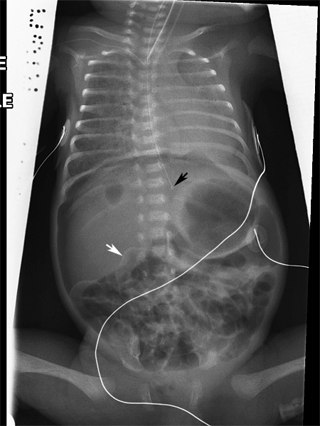
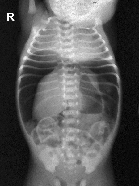
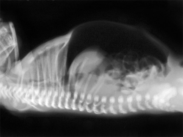
Pneumoperitoneum
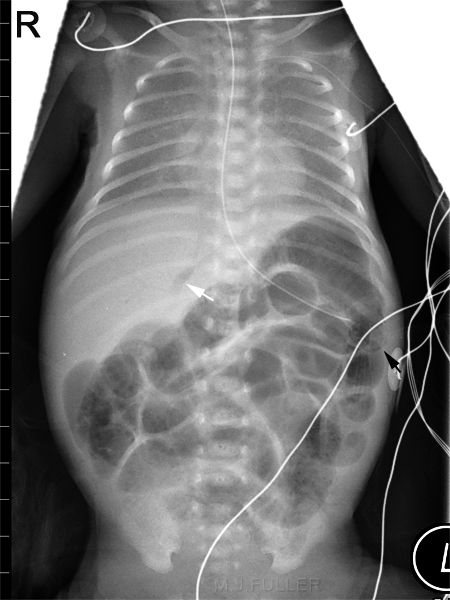
Necrotising Enterocolitis (NEC)
(Created page with "<div class="WPC-editableContent"><br/><br/><b><font size="4">Pneumoperitoneum</font></b><br/><blockquote><table align="bottom" cellpadding="3" class="WPC-edit-style-grid1 WPC-...") |
(No difference)
|
Latest revision as of 17:16, 11 November 2020
Pneumoperitoneum
Necrotising Enterocolitis (NEC)
Intramural gas and Portal-venous GasDefinition
- Defined as an idiopathic coagulation necrosis and inflammation of the intestine in a neonatal patient.
Onset
- The age of onset is highly variable but rarely occurs in the first three days of life.
- The lowest GA (24-28 weeks) tend to develop NEC after the second week of lifeIntermediate GA (29-32 weeks) develop it within 1-3 weeks
- Term infants or >32 weeks tend to develop it in the first week of life.
Bell’s staging criteria
- Stage I (suspected NEC)
- Stage II (definite NEC)
- Stage III (advanced NEC, severely ill)
- IIIA (without perforation)
- IIIB (with perforation)
Radiological Appearance•Generalized bowel distention (earliest sign)•Pneumatosis Intestinalis•Pneumoperitoneum•Large distended immobile loop on repeated x-rays(persistant loop sign) (may indicate a gangrenous loop of bowel)
•Gasless abdomen (perforation and peritonitis)•Portal venous air
<a class="external" href="http://www.ohsu.edu/ohsuedu/academic/som/pediatrics/clerkships/upload/Necrotizing_enterocolitis_5_2005.ppt." rel="nofollow" target="_blank">
Necrotizing enterocolitis Charlene Crichton, MD</a>
Transient Portal venous Air
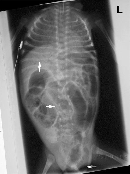
This baby has portalvenous gas (top arrow) intramural gas(middle arrow) and an inguinal hernia (bottom arrow)
" It is only in the neonate that visible intramural gas, as in Necrotising enterocolitis, is survivable (case report). Intramural gas in the adult usually marks infarction, necrosis and an indication for resection" <a class="external" href="http://myweb.lsbu.ac.uk/dirt/museum/p7-abdo.html" rel="nofollow" target="_blank">http://myweb.lsbu.ac.uk/dirt/museum/p7-abdo.html</a>
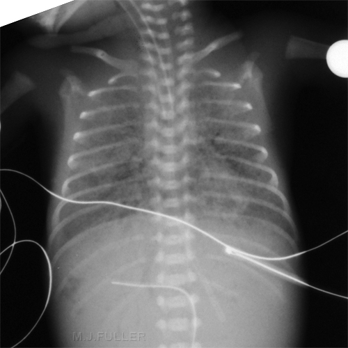
You should not rely on your ability to establish which is the correct left-right orientation of the image after it is taken. Pathology and patient rotation can make this very difficult. This patient's faintly visible left ventricle and nasogastric tube help, but what if the patient had situs inversus?
The UVC tip has tracked into the liver and there appears to be faint portal venous gas.
Air in portal venous branches can be associated with umbilical venous catheter insertion. Inconsequential transient portal venous air can be seen immediately after umbilical venous catheter insertion and should not necessarily be attributed to necrotizing enterocolitis.
<a class="external" href="http://www.ajronline.org/cgi/content/full/180/4/1147" rel="nofollow" target="_blank">Alan E. Schlesinger1, Richard M. Braverman1 and Michael A. DiPietro2 </a><a class="external" href="http://www.ajronline.org/cgi/content/full/180/4/1147" rel="nofollow" target="_blank">AJR 2003; 180:1147-1153</a>
Malrotation with Volvulus
"Malrotation of the intestines results when the intestinal rotation and fixation that occurs during pregnancy fails to occur. This normally happens in the 4th and 12th weeks of fetal life. In the 4th fetal week, the entire bowel is basically a straight tube with the superior mesenteric artery (SMA). During the course of pregnancy, the bowel rotates in place to the left of the SMA at the ligament of Treitz" <a class="external" href="http://www.pedisurg.com/PtEduc/Malrotation.htm" rel="nofollow" target="_blank">Texas Pediatric Surgical Associates</a>
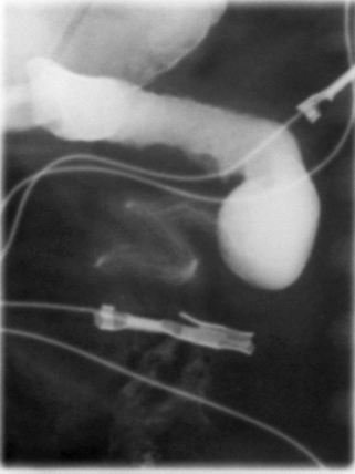
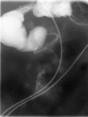
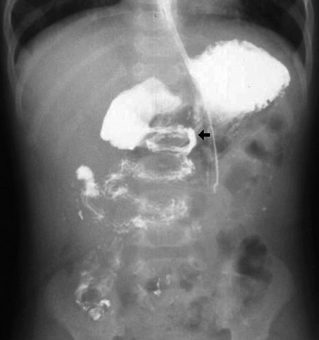
Malrotation of the gut has a characteritic "corkscrew" appearance on a barium study. It is important that the barium column is observed to cross the spine as it passes through the deuodenum.
"Rotational abnormality of the gut can be entirely asymptomatic but in other cases the sequelae are catastrophic when Ladd’s band or volvulus leads to obstruction. Failure of normal embryological bowel rotation leaves the superior mesenteric vein (SMV) anterior and to the left of the superior mesenteric artery (SMA) as opposed to its normal position to the right of the artery. When volvulus occurs the entire gut twists or corkscrews around the SMA and leads to vascular compromise. This may be complicated with necrosis, perforation and gangrene.
The findings are quite variable on a plain abdominal radiograph. They can range from a normal appearing abdomen through one suggesting a gastric outlet to one suggesting small bowel obstruction. The ultimate picture depends on the degree of volvulus (i.e. number of twists of the bowel ), duration of volvulus and the small bowel involved.
Contrast studies with Barium confirm the level of obstruction which is usually towards the third and fourth parts of duodenum, and often demonstrates the compressing band. The small bowel may show characteristic "corkscrew" or "spiral" pattern. " <a class="external" href="http://www.kem.edu/dept/radiology/case34_01.htm" rel="nofollow" target="_blank">http://www.kem.edu/dept/radiology/case34_01.htm</a>
<a class="external" href="http://www.hawaii.edu/medicine/pediatrics/pemxray/v3c17.html" rel="nofollow" target="_blank">Source: Loren G. Yamamoto, MD, MPH </a>
<a class="external" href="http://www.hawaii.edu/medicine/pediatrics/pemxray/v3c17.html" rel="nofollow" target="_blank">Kapiolani Medical Center For Women And Children </a>
<a class="external" href="http://www.hawaii.edu/medicine/pediatrics/pemxray/v3c17.html" rel="nofollow" target="_blank">University of Hawaii John A. Burns School of Medicine</a><a class="external" href="http://www.hawaii.edu/medicine/pediatrics/pemxray/v3c17.html" rel="nofollow" target="_blank"> </a><a class="external" href="http://www.hawaii.edu/medicine/pediatrics/pemxray/v3c17.html" rel="nofollow" target="_blank"> </a>
<a class="external" href="http://www.hawaii.edu/medicine/pediatrics/pemxray/v3c17.html" rel="nofollow" target="_blank">Source: Loren G.</a><a class="external" href="http://www.hawaii.edu/medicine/pediatrics/pemxray/v3c17.html" rel="nofollow" target="_blank">Yamamoto, MD, MPH </a>
<a class="external" href="http://www.hawaii.edu/medicine/pediatrics/pemxray/v3c17.html" rel="nofollow" target="_blank">Kapiolani Medical Center For Women And Children </a>
<a class="external" href="http://www.hawaii.edu/medicine/pediatrics/pemxray/v3c17.html" rel="nofollow" target="_blank">University of Hawaii John A. Burns School of Medicine</a>This study from the University of Hawaii gives a better impression of the malrotated anatomy
Typical barium study appearance:
o Duodenojejunal junction (ligament of Treitz) located lower than duodenal bulb and to the right of expected position
o Spiral course of midgut loops = "apple-peel / twisted ribbon / corkscrew" appearance (in 81%)
