Neck
Jump to navigation
Jump to search
Neck Surface Anatomy | Other pages of interest |
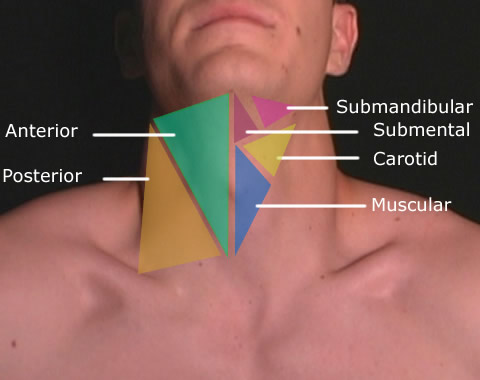
Triangles of the Neck
| | Anatomists use the term triangles of the neck to describe the divisions created by the major muscles in the region. The most commonly used are the Anterior and Posterior triangles. The anterior triangle of the neck is bounded, in front, by the mid line of the neck, behind by the anterior margin of the sternocleidomastoideus, its base is formed by the lower border of the body of the mandible. The posterior triangle of the neck has borders of the posterior border of the sternocleidomastoid muscle, middle third of the clavicle and the anterior border of the trapezius muscle. The carotid triangle is a portion of the anterior triangle of the neck where the common carotid artery bifurcates. The submental triangle contains two lymph glands and the submental lymph nodes. The submandibular triangle contains the submaxillary gland and part of the parotid gland. The muscular triangle is bounded, in front by the median line of the neck from the hyoid bone to the sternum, behind by the anterior margin of the Sternocleidomastoideus and above by the superior belly of the omohyoideus. |
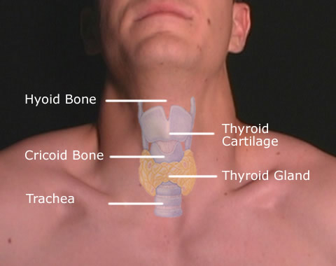
Neck - Anterior Surface
| | The Adam's apple clinically known as the laryngeal prominence is a lump/ protrusion formed by the angle of the thyroid cartilage surrounding the larynx. This is an often used centering point in radiography for the cervical spine AP view. The thyroid is one of the largest endocrine glands in the body. This gland is found in the anterior neck inferior to the mandible. The thyroid controls metabalism in the body and is one of the most sensitive areas of the body to radiation. |
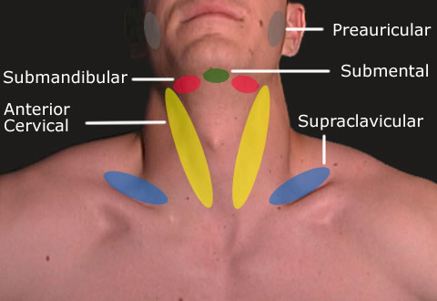
Superficial Neck Lymph Nodes
| | Lymph nodes are components of the lymphatic system and there are approximately 300 lymph nodes in the neck. Nodes act as filters, with an internal honeycomb of reticular connective tissue filled with lymphocytes that collect and destroy bacteria and viruses. When the body is fighting an infection, lymphocytes multiply rapidly and produce a characteristic swelling of the lymph nodes. The location of the Preauricular, Submandibular, Submental, Anterior Cervical and Supraclavicular superficial node chains are shown below. |
Go Back to Surface Anatomy Index


