Modified Lateral Thoracic Spine Technique
Introduction
One of my radiography colleagues performs a novel lateral thoracic spine radiographic technique that is as clever as it is effective. Whilst this might not be an acceptable routine technique in your institution, you may find it a useful tool to have in your radiography toolbox.
TechniqueThe technique basically combines the lateral thoracic spine positioning technique with the swimmers position for demonstrating the cervico-thoracic junction. The patient is positioned initially on the X-ray table in the lateral position. The patient's hands would normally be placed together in front of the patient's head. The variation in technique comes from modifying the position of the arm that is away from the X-ray table. Ask the patient to place this arm behind his/her back and roll this shoulder posteriorly until it is clear of the thoracic spine. This requires some flexibility from the patient. If the patient does not have a good range of shoulder movement, the patient will have to be rolled too far 'off lateral' to clear this shoulder off the thoracic spine.
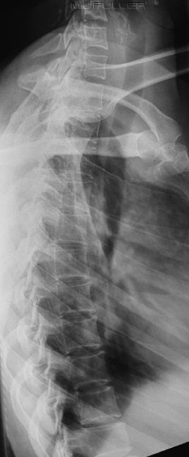
You should achieve a result something like this-
A breathing technique is highly desirable.
One of the disadvantages of this technique is that it will often result in the patient rolling off true lateral. This is undesirable, but in practice it is unlikely that a lesion would be missed that would have been demonstrated on a more conventionally positioned patient. On the contrary, upper thoracic spine lesions have been demonstrated that might otherwise have been missed. Coned views of lesions of interest can be performed as a supplementary view.
The desire to perform a perfect lateral is part of the standardisation of positioning in radiography and is a worthy objective. However, I would argue that there is a risk of doctrinaire thinking on this issue.
This patient presented to the Emergency Department and was referred for thoracic spine radiography with special emphasis on the upper thoracic spine and thoraco-cervical junction. The image is a result of positioning and exposure technique rather than something that can only be achieved with digital equipment. This is a plain film radiograph not a digital image.
Case Study
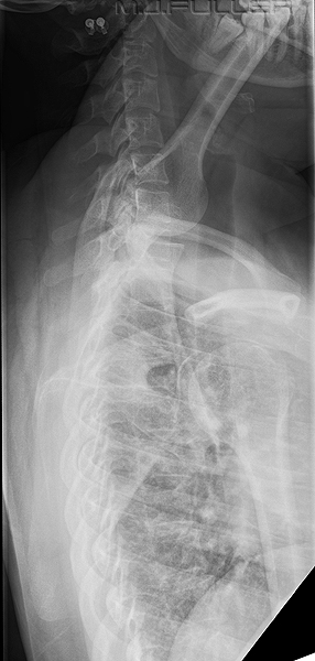
This patient has sustained an injury to her upper thoracic vertebra. The patient has been referred for serial lateral thoracic spine imaging for ongoing assessment. Compare the two radiographic techniques. The whole of the thoracic and cervical spine anatomy have been included at the request of the referring clinician. These images were taken using a CR system.
The patient is positioned in the lateral position with arms forward- one up and one down. A kVp has been set by the radiographer and the automatic exposure device has determined the mA and time values.
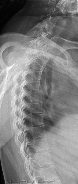
This is the modified lateral position as described above. In addition, an exposure time of 1.6 seconds has been employed and the patient has continued breathing during the exposure. An aluminium filter has been positioned on the LBD to coincide with the cervical spine.
The combination of modified patient positioning, breathing technique and the use of an aluminium filter has resulted in a dramatic improvement in image quality.
....back to the applied radiography home page here


