Maisonneuve Fracture
Introduction
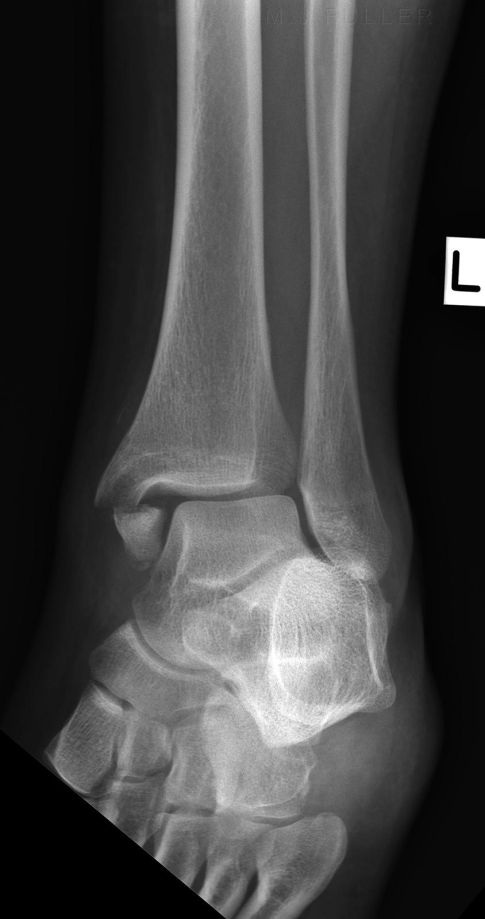
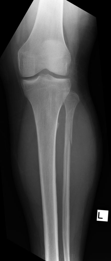
Maisonneuve fractures are commonly missed clinically and radiographically. The proximal fibular fracture is often not appreciated because of the distracting ankle injury. Radiographers should be aware of the Maisonneuve fracture, its mechanism of injury and the potential for an asymptomatic proximal fibular fracture. When a syndesmosis injury is demonstrated on ankle radiography, the radiographer should consider the possibility of a Maisonneuve fracture and undertake supplementary views to demonstrate the entire lower leg.
Definition
The Maisonneuve fracture consists of a proximal fibular fracture with associated ligament disruption or medial malleolar ankle fracture. <a class="external" href="http://www.ispub.com/journal/the_internet_journal_of_orthopedic_surgery/volume_4_number_1_38/article/maisonneuve_fracture_associated_with_a_posterior_malleolar_fracture_a_case_report.html" rel="nofollow" target="_blank">(http://www.ispub.com/journal/the_internet_journal_of_orthopedic_surgery/volume_4_number_1_38/article/maisonneuve_fracture_associated_with_a_posterior_malleolar_fracture_a_case_report.html)</a>
The Maisonneuve fracture of the fibula is often considered one of the most unstable ankle injuries. This is because of the presumed disruption of all the syndesmotic ligaments and the interosseous membrane from the ankle joint to the level of the fibula fracture. <a class="external" href="http://www.ortopediavirtual.com.br/docs/Maisonneuve.pdf" rel="nofollow" target="_blank">KEITH DOUGLAS MERRILL, M.D. The Maisonneuve Fracture of the Fibula</a>
Pathophysiology
The primary motion of the ankle at the true ankle joint (tibiotalar joint) is plantarflexion and dorsiflexion. Inversion and eversion occur at the subtalar joint.
Excessive inversion stress is the most common cause of ankle injuries for 2 anatomic reasons.
- First, the medial malleolus is shorter than the lateral malleolus, allowing the talus to invert more than evert.
- Second, the deltoid ligament stabilizing the medial aspect of the ankle joint offers stronger support than the thinner lateral ligaments. As a result, the ankle is more stable and resistant to eversion injury than inversion injury. However, when eversion injury occurs, there is often substantial damage to bony and ligamentous supporting structures and loss of joint stability.
<a class="external" href="http://emedicine.medscape.com/article/824224-overview" rel="nofollow" target="_blank">quoted from http://emedicine.medscape.com/article/824224-overview</a>
Anatomy
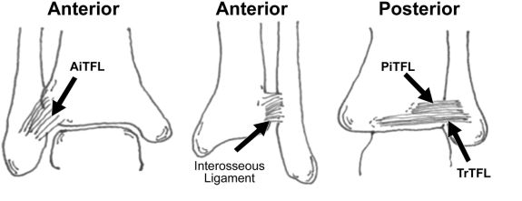
Ankle Syndesmosis
<a class="external" href="http://radiology.rsna.org/content/242/1/225/F1.expansion" rel="nofollow" target="_blank">
Chronic Tibiofibular Syndesmosis Injury of Ankle: Evaluation with Contrast-enhanced Fat-suppressed 3D Fast Spoiled Gradient-recalled Acquisition in the Steady State MR Imaging</a>Ankle Syndesmosis
- syndesmosis is made up of 4 ligaments which stabilize the distal tibiofibular syndesmosis:
- anterior-inferior tibiofibular ligament (AITFL)
- interosseous ligament,
- posterior- inferior tibiofibular ligament (PiTFL)
- inferior transverse tibiofibular ligament (TrTFL)
<a class="external" href="http://www.wheelessonline.com/ortho/syndesmotic_injuries_of_the_ankle" rel="nofollow" target="_blank">http://www.wheelessonline.com/ortho/syndesmotic_injuries_of_the_ankle</a>
Case 1
... back to the Applied Radiography home page


