Lunate and Perilunate Dislocations
Lunate and perilunate dislocations following trauma to the wrist are uncommon but not rare. These injuries are unfortunately missed too often and present considerable management problems when diagnosis is delayed.
Anatomy
Normal Variants
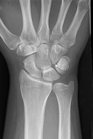
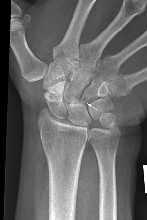
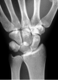
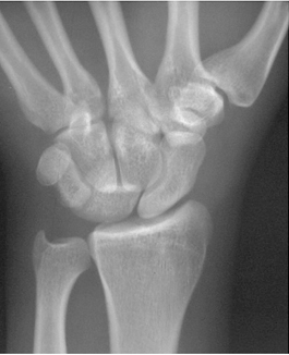
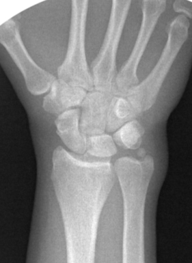
This is a type I lunate. Note that the lunate does not articulate directly with the hamate. This is a type II lunate- the hamate articulates directly with the lunate.
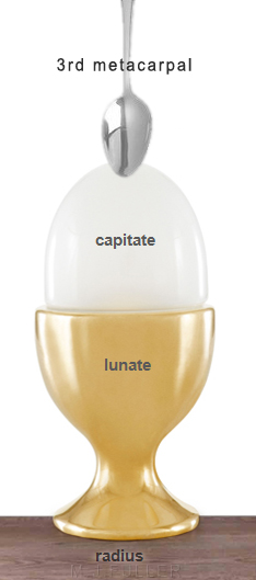
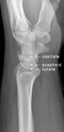
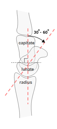
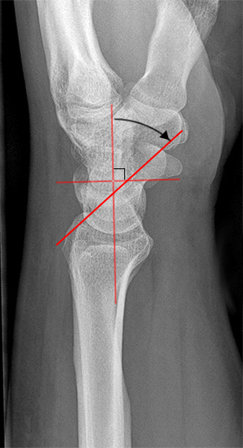
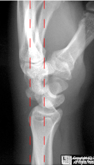
The Distal Radius, Scaphoid, Capitate, Lunate and Metacarpal RelationshipWhen you review your wrist radiography in a trauma setting, it is important to consider whether you can see a normal relationship between the distal radius, lunate, scaphoid and capitate. It is fortunate that the lunate, scaphoid and capitate are often the easiest to visualise- all three have a smooth proximal convex cortical margin and they almost always occur in the same order.
Lunate Dislocation or Perilunate Dislocation?
Perilunate Dislocation Radiological Appearance
- 4 types of perilunate dislocations
1 transscaphoid-perilunate
2 perilunate
3 transradial-styloid
4 transscaphoid-trans-capitate-perilunar
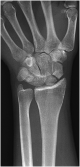
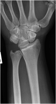
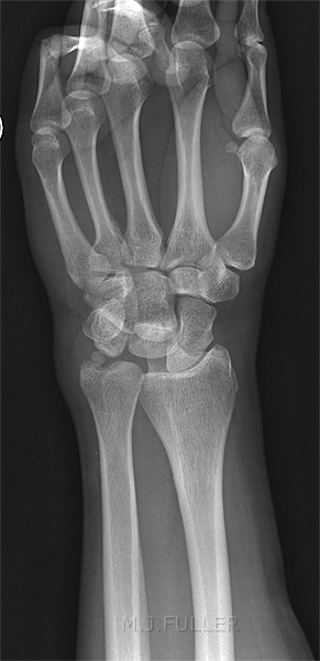
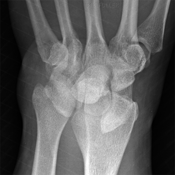
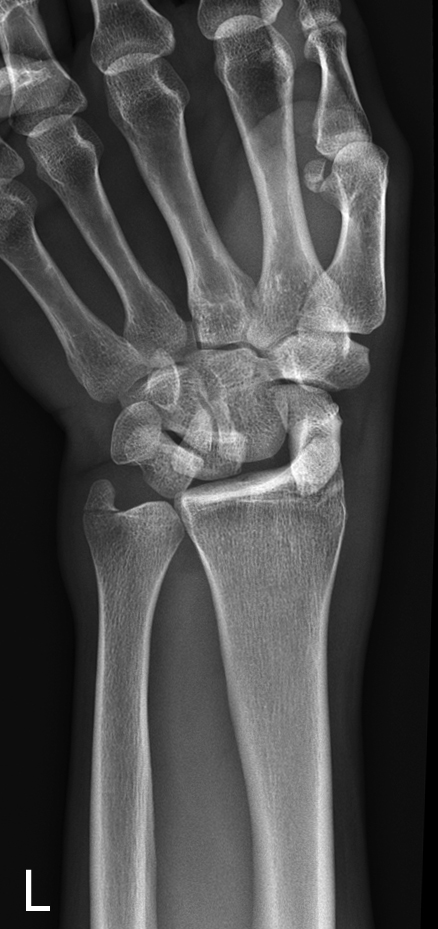
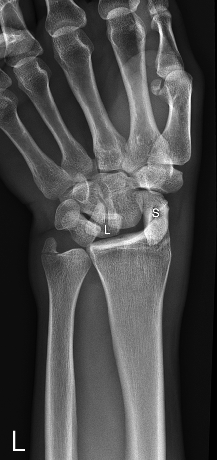
This 32 year old man presented to the Emergency Department after falling from a ladder. He was referred for an extensive list of radiographic examinations including left wrist. This is the first image from this particular trauma series. (in cases of serious trauma, the wrist imaging would not be a high priority)
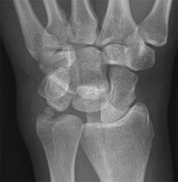
There is loss of parallelism of the carpal bones. The ulnar styloid suggests a normal variant or old injury. There is loss of the normal proximal and distal carpal arc lines
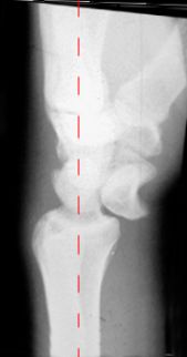
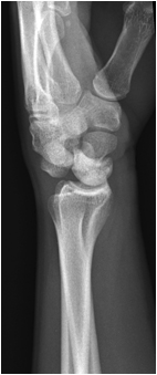
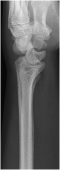
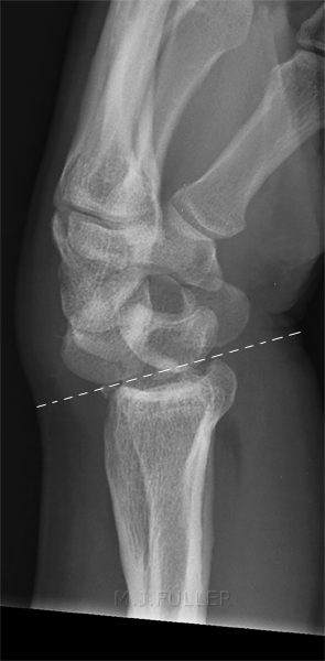
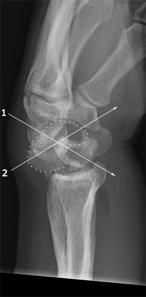
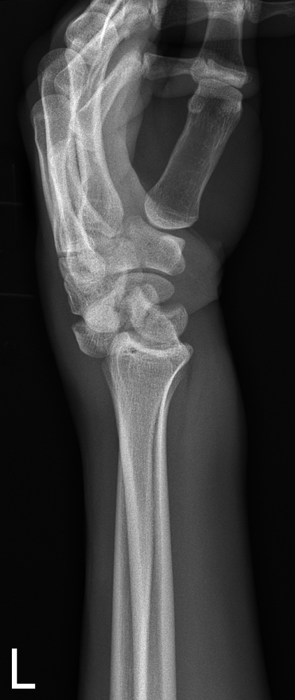
The loss of normal carpal arcs was noted by the radiographer who proceeded to perform a lateral wrist view.
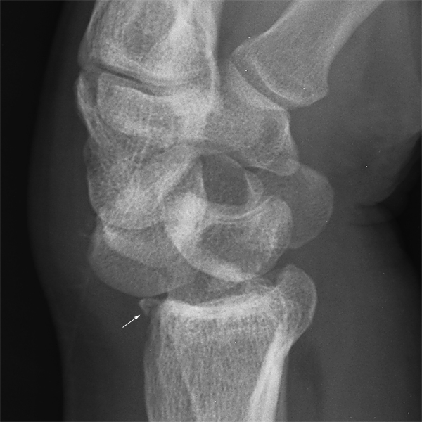
The radiographer paid particular attention to the integrity of the scaphoid.
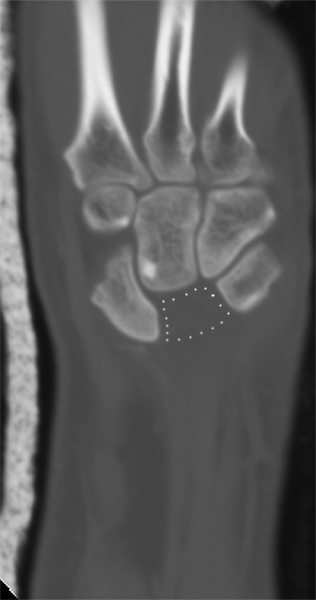
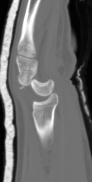
The patient proceeded to CT followed by the operating theatre for reduction of the carpal bone dislocation.
This is the post-reduction image from the mobile image intensifier. Comment
This case demonstrates a thoughtful approach to the radiographic demonstration of this patient's carpal injury. The radiographer recognised the carpal dislocation from the PA image. The radiographer was mindful of the importance of obtaining a good lateral projection. Equally, the radiographer paid careful attention to the imaging of the scaphoid (given the association between this carpal dislocation and scaphoid fracture). Importantly, the surgical approach in cases where a scaphoid fracture is demonstrated is different to a simple closed reduction of a perilunate dislocation.
The radiographer also noted the negative palmar tilt and considered the possibility of an old wrist fracture. This was confirmed by the patient and this information was relayed in person by the radiographer to the reporting radiologist.
This case supports the contention that trauma radiography is a specialised area of plain film radiography. In addition to all of the above, the radiographer relayed the findings in person to the referring doctor in the ED. This facilitated a timely undertaking of orthopaedic review and subsequently CT imaging of the wrist and surgical reduction. This type of personal communication helps to engender a co-operative relationship, sense of common purpose, and camaraderie between the radiographer and the referring doctor.
Finally, I would suggest that the satisfaction experienced by the radiographer in successfully completing this examination would be vastly different to a radiographer who simply undertook AP and lateral views of the patient's wrist and was unaware that the patient had sustained a significant wrist injury.
Case 2
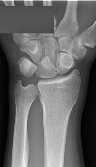
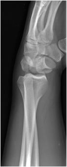
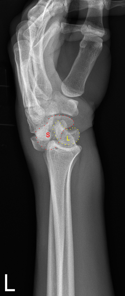
This 28 year old male presented to the Emergency Department following unknown trauma with a painful left wrist which appeared grossly abnormal. He was referred for left wrist radiography.
There is a posterior perilunate dislocation
The lunate appears to be normally aligned
Case 3
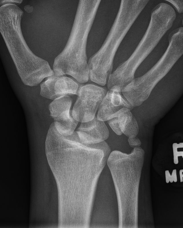
This 25 year old male presented to the Emergency Department following a fall. He was examined and found to have a very painful right wrist. He was referred for right wrist radiography.
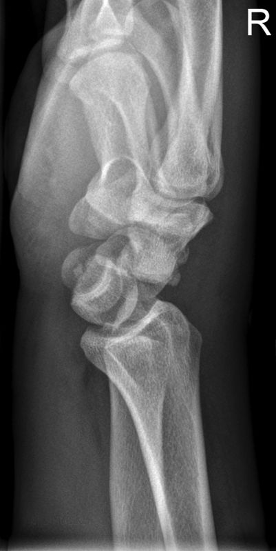
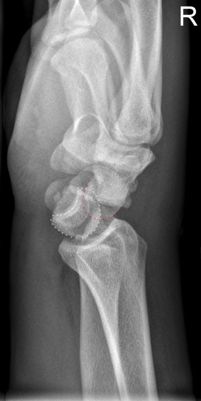
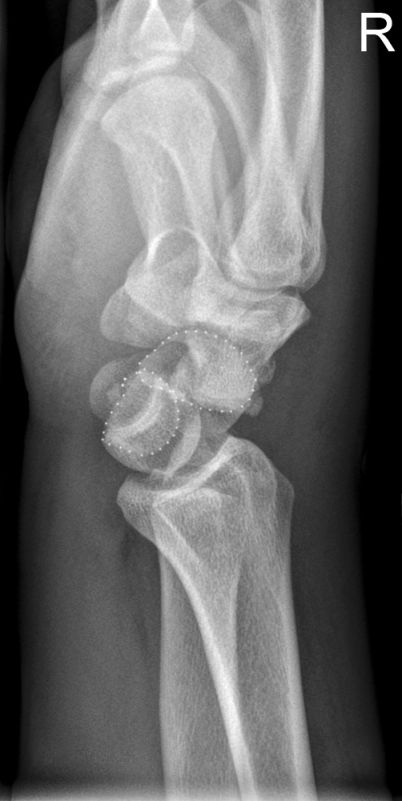
There is a scaphoid cortical irregularity and fracture line suggesting a scaphoid fracture. The lunate is flexed (volar rotation)- this is referred to as a piece-of-pie sign.
There is discontinuity of Gilula's second arc at the lunate.
The distance between the proximal cortical border of the capitate and the corresponding articular surface of the lunate is abnormally large (parallelism)
The appearances suggest a transcaphoid perilunate dislocation
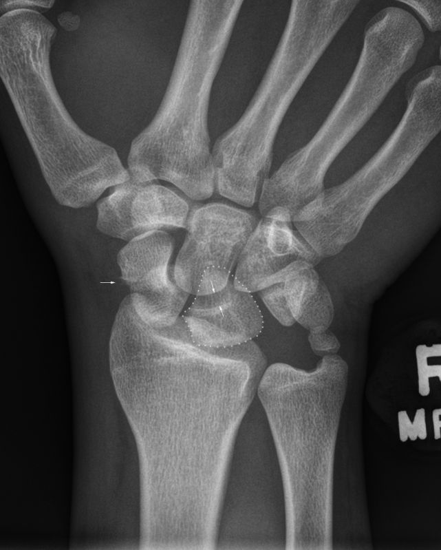
Old fracture of the ulnar styloid is noted.The lunate is outlined.
The distance between the lunate and capitate is abnormally large (parallelism)The lunate is displaced in a volar direction. There is a bony fragment volar to the lunate which is a fragment of the scaphoid. The proximal capitate is positioned dorsal to the lunate.
The lunate is outlined in white
The proximal capitate is outlined in red.The two fragments of the fractured scaphoid are outlined in white. <embed allowfullscreen="true" height="350" src="http://widget.wetpaintserv.us/wiki/wikiradiography/widget/youtubevideo/1dc9e736f82c338d9c00123250d7f636b249f00c" type="application/x-shockwave-flash" width="425" wmode="transparent"/>
If you click on the you tube logo you can watch this video at full resolution. You will need to select 720p and the fit-to-screen buttons to see the video in full resolution.Select the Esc key on your keyboard to return to normal viewing.
<embed allowfullscreen="true" height="350" src="http://widget.wetpaintserv.us/wiki/wikiradiography/widget/youtubevideo/3e672e0c46a6806de50ef9608df254874a31c437" type="application/x-shockwave-flash" width="425" wmode="transparent"/> If you click on the you tube logo you can watch this video at full resolution. You will need to select 720p and the fit-to-screen buttons to see the video in full resolution. Select the Esc key on your keyboard to return to normal viewing.
<embed allowfullscreen="true" height="350" src="http://widget.wetpaintserv.us/wiki/wikiradiography/widget/youtubevideo/bbc6c84bd3333de1d8f3ae7ac2d43d9fbc3f8a43" type="application/x-shockwave-flash" width="425" wmode="transparent"/> If you click on the you tube logo you can watch this video at full resolution. You will need to select 720p and the fit-to-screen buttons to see the video in full resolution. Select the Esc key on your keyboard to return to normal viewing.
... back to the Home page
... back to the Applied Radiography home page
