IntroductionLumbar spine radiography is an increasingly uncommon examination in centres with CT and MRI imaging. Despite this, there is still a case for plain film imaging of the lumbar spine in elderly patients, in cases of significant trauma, and/or when there is a possibility of infection or tumour.
AP/PA Lumbar SpineAP or PA?
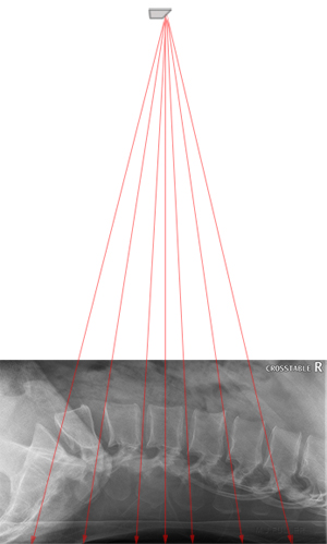
The PA projection of the lumbar spine has the advantage of matching the divergent X-ray beam with the patient's lumbar lordosis. Conversely, the AP projection of the lumbar spine has the divergent beam working against the patient's lumbar lordosis.
AP Lumbar Spine Projection
(patient supine)
 | PA Lumbar Spine Projection
(patient prone)
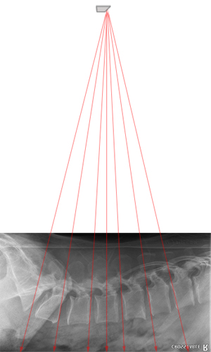 |
| The AP lumbar spine projection tends to demonstrate the central disc space only in profile. This effect can be minimised by flexing the hips and knees to reduce the lumbar lordosis. | The PA projection of the lumbar spine tends to demonstrate all of the intervertrebral dics in profile |
The PA projection of the lumbar spine has the advantage of matching the divergent X-ray beam with the patient's lumbar lordosis. Conversely, the AP projection of the lumbar spine has the divergent beam working against the patient's lumbar lordosis.
Exposure technique
The breathing exposure technique is usually associated with lateral thoracic spine radiography. The same technique can be employed for all torso spine plain film imaging.
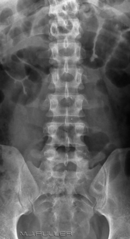
AP Lumbar Spine Projection
(short exposure time)
 | AP Lumbar Spine Projection
(breathing technique)
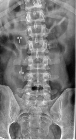 |
| This AP lumbar spine image was produced using the automatic exposure device and a short exposure time. The soft tissues of the abdomen distract from the image of the bony anatomy | This is a repeat view using a breathing technique. There is improved demonstration of the bony anatomy. |
Lateral Lumbar SpineHorizontal Ray Technique
The horizontal ray lateral technique is commonly employed in patient who are unable to rotate into the lateral position and/or in patients who have a possibility of an unstable spinal injury.
Horizontal Ray Lateral Lumbar Spine
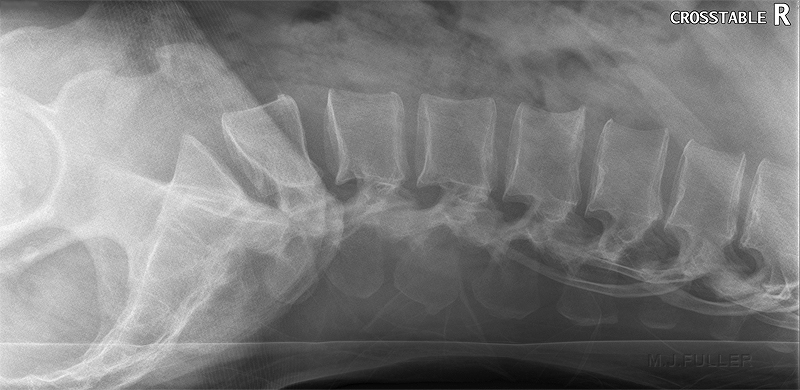 |
| This is a horizontal ray lateral lumbar spine image in a patient who was involved in a motor vehicle accident. A long exposure breathing technique has been employed. The results are comparable with a conventional rolled lateral lumbar spine technique. |
Exposure technique
The breathing exposure technique is usually associated with lateral thoracic spine radiography. The same technique can be employed for all torso spine plain film imaging.
AP Lumbar Spine Projection
(short exposure time)
 | AP Lumbar Spine Projection
(breathing technique)
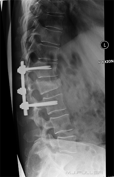 |
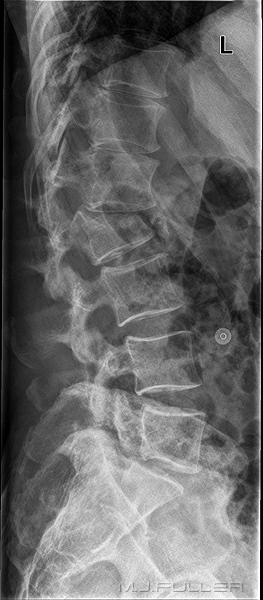
There is a crush fracture of the body of L2. The patient was referred for a surgical assessment and received pedicle screws to stabilise the fracture.
The radiographer set the kVp at 85 and the automatic exposure device determined the exposure mA and time. The exposure was made on arrested respiration. Note that there are bowel gas and diaphragmatic shadows overlying the spine. | The patient returned to the X-ray department following spinal surgery for post-operative imaging. The radiographer selected the following manual exposure factors
The long exposure time has resulted in blurring of the bowel gas, ribs and diaphragm. The spinal bony anatomy remains sharp. The patient was asked to hold still but remain breathing during the exposure. This technique should not result in an increase in the mAS used- the mA is reduced to match the increased exposure time. |
Oblique Lumbar SpineRadiographic Technique
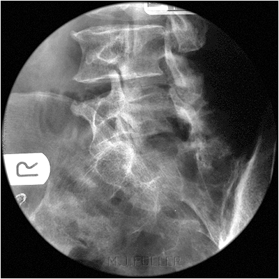 | 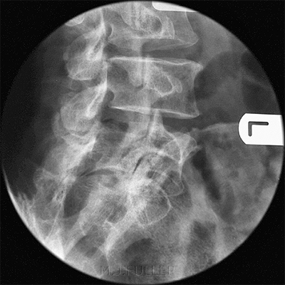 |
This is the RPO or LAO position to show the lower lumbar vertebra.
Film/screen technique with round cone | This is the LPO or RAO position to show the lower lumbar vertebra
Film/screen technique with round cone |
Centre Point for PA Technique
| Incorrect | Correct |
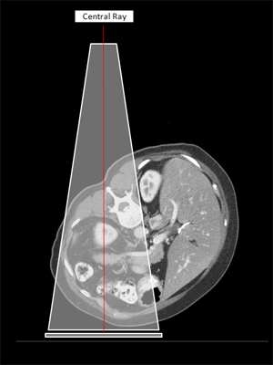 | 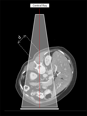 |
| This is a trap that you might fall into the first time you attempt an oblique lumbar spine in the prone/PA oblique position. The beam is centred to what appears to be the spine based on surface anatomy | This is the correct positioning. Centre onto the raised side by at least an inch (d = 3.5cm). The exact centring point will depend on the patient's body habitus and degree of obliquity |
Should you Include all of the Lumbar Vertebrae on Oblique Views?
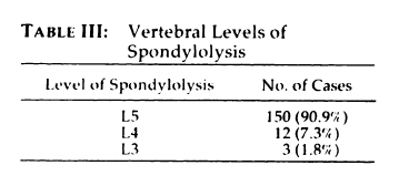
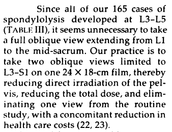
Libson et al (Radiology 1984; 151: 89-90) reported the following
To take the authors argument even further, the diagnostic yield from including L3 will be very small and arguably not worth the cost in terms of patient radiation dose.
...back to the
Applied Radiography home page


