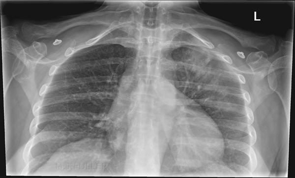
Left Upper Lobe Consolidation
The Left Upper Lobe (LUL) is a relatively uncommon site for consolidation. The diagnosis of a subtle LUL consolidation can be very tricky on the PA/AP view image and can be relatively easy on the lateral image.
The Meaning of the Term Consolidation
One of the unfortunate aspects of the term consolidation is that its meaning can be different depending on who is using the term. When a clinician uses the term consolidation he/she is usually referring to a consolidation associated with acute pneumonia. Thus, the term consolidation and pneumonia have very similar meanings and are almost used interchangeably.
Strictly speaking, the term consolidation does not imply any particular aetiology or pathology. Acute pneumonia is the commonest cause but not the only cause of consolidation. (other causes include chronic pneumonia, pulmonary oedema and neoplasm). Thus when a radiologist has reported a chest X-ray examination and notes the presence of consolidation he/she is simply stating that some of the long airspace has been replaced by a fluid.
Notes on Consolidation
- Refers to fluid in the airspaces of the lung
- Consolidation may be complete or incomplete
- The distribution of the consolidation can vary widely. A consolidation could be described as “patchy”, “homogenous”, or generalised”.
- A consolidation may be described as focal or by the lobe or segment of lobe affected
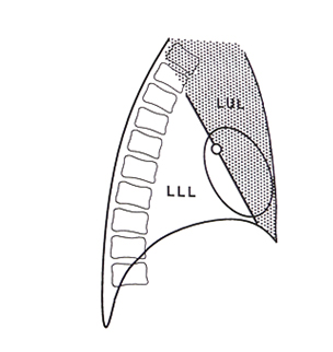
The Left Upper Lobe (LUL) Anatomy
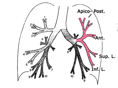
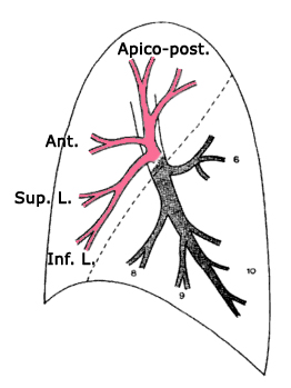
adapted from <a class="external" href="http://books.google.com.au/books?id=Bif0zpmEWtAC" rel="nofollow" target="_blank">By Fred W. Wright Radiology of the Chest and Related Conditions: Together with an Extensive Illustrative Collection of Radiographs CRC Press, 2002</a>On the left there is no middle lobe; the anatomical equivalent region corresponding to the right middle lobe is known as the lingula, and like the RML, is also composed of two segments. Unlike their counterparts on the right however, the segments are stacked one on top of another, rather than side.
<a class="external" href="http://lib.cpums.edu.cn/jiepou/tupu/atlas/www.vh.org/adult/provider/radiology/LungAnatomy/RightLung/RtLungSegAnat.html" rel="nofollow" target="_blank">http://lib.cpums.edu.cn/jiepou/tupu/atlas/www.vh.org/adult/provider/radiology/LungAnatomy/RightLung/RtLungSegAnat.html</a>.
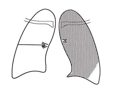
Note that upper lobe pathology could appear very low on a chest X-ray image. The upper lobe is the anterior lobe as much as it is the upper lobe.
adapted from <a class="external" href="http://books.google.com.au/books?id=Bif0zpmEWtAC" rel="nofollow" target="_blank">By Fred W. Wright Radiology of the Chest and Related Conditions: Together with an Extensive Illustrative Collection of Radiographs CRC Press, 2002</a>
Plain Film Appearances of Lung Consolidation
Radiological appearances common to all lobes are:
1.Abnormal lung opacity2.Increase in the size and number of lung markings3.Loss of clarity of the diaphragm on the AP and/or lateral views4.Loss of clarity of the heart border on the AP and/or lateral views5.Air bronchogram lines6.Loss of the normal darkening inferiorly of the thoracic vertebral bodies on the lateral view7.Opacification of the lung behind the heart shadow or below the diaphragms
Case 1
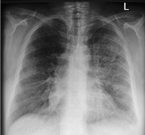
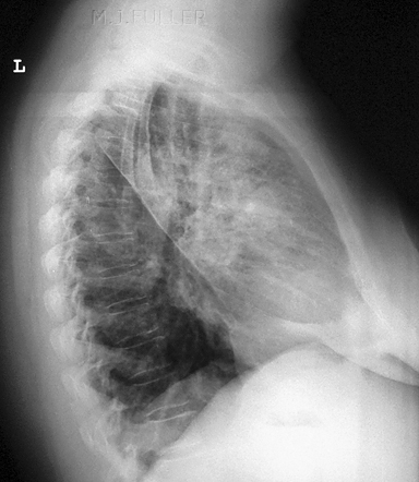
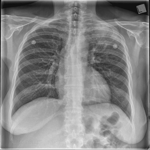
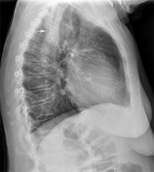
There is increased abnormal opacity seen within the apex of the left lung. This is likely to represent consolidation within the LUL.
Benjamin Felson (<a class="external" href="http://www.amazon.com/Chest-Roentgenology-Benjamin-Felson/dp/0721635911/ref=sr_1_2?ie=UTF8&s=books&qid=1252240078&sr=1-2" rel="nofollow" target="_blank">Chest Roentgenology, W.B. Saunders, 1973, p22</a>) notes that "A radiopacity involving the extreme apex of the lung is almost invariably situated in the apical segment of the upper lobe".There is consolidation within the LUL confirmed on the lateral chest image. The opacity may appear deceptively dense because of the superimposed densities. An apical lordotic view was performed to see if the pathology could be better demonstrated.
... back to the Applied Radiography home page
