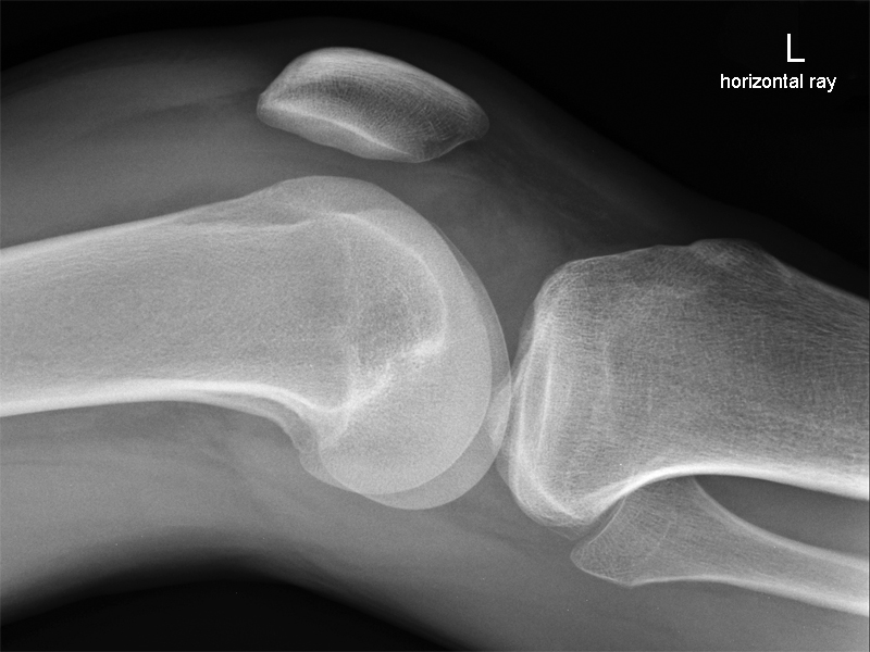
Lateral Knee Radiography
Indications for Knee RadiographyLateral knee radiography commonly raises a number of questions:
- is there a reliable technique for lateral knee radiography?
- how do I correct a lateral knee malposition?
- when is it malpositioned enough to warrant a repeat?
This page attempts to answer these and other lateral knee radiography questions
Ottawa rules
State that a knee X-ray is only required for patients with knee injuries with any of the following:
- Age 55 or over.
- Isolated tenderness of the patella (no bone tenderness of the knee other than the patella).
- Tenderness at the head of the fibula.
- Inability to flex to 90 degrees.
- Inability to weight bear both immediately and in the casualty department (ie, 4 steps – unable to transfer weight twice onto each lower limb regardless of limping).
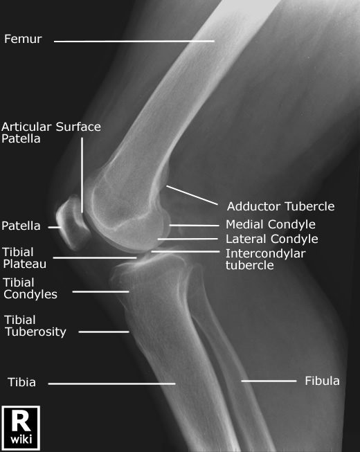
Anatomy<a class="external" href="http://www.imageinterpretation.co.uk/knee.html" rel="nofollow" target="_blank">Image Interpretation Course</a>
<a class="external" href="http://www.imageinterpretation.co.uk/knee.html" rel="nofollow" target="_blank">by Heidi Gable DCR(R) PgCert</a>
<a class="external" href="http://www.imageinterpretation.co.uk/knee.html" rel="nofollow" target="_blank">http://www.imageinterpretation.co.uk/knee.html</a>
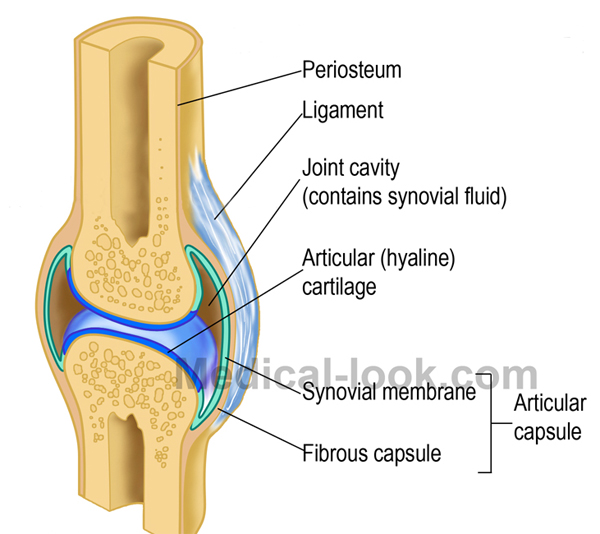
Synovial Joint Synovium or capsule? Synovial CavityThe terms synovium and capsule do appear to get confused- they are not interchangeable.
- synovial cavity is deepest layer of joint capsule
- although synovium membrane is attached all around, above to articular
margins of femur and below to articular margins of tibia, it is not
everywhere coextensive with the capsule or ligament and tendons- because of folds of synovial membrane, synovial cavity is not a simple,
short, cylindrical cavity- behind and above the patella, it is single cavity that is usually
continuous above w/ suprapatellar bursa between the tendon of
quads & femur;- cruciate ligaments, meniscii, & infrapatellar fat pad are
outside the synovial cavity;<a class="external" href="http://www.wheelessonline.com/ortho/synovium_of_the_knee" rel="nofollow" target="_blank">http://www.wheelessonline.com/ortho/synovium_of_the_knee</a>
- Synovium is very variable but often has two layers.
- The outer layer, or subintima, can be of almost any type: fibrous, fatty or loosely "areolar".
- The inner layer, or intima, consists of a sheet of cells thinner than a piece of paper.
- Where the underlying subintima is loose the intima sits on a pliable membrane, giving rise to the term synovial membrane. This membrane, together with the cells of the intima, provides something like an inner tube, sealing the synovial fluid from the surrounding tissue (effectively stopping the joints being squeezed dry when subject to impact, such as running).
- The intimal cells are of two types, fibroblasts and macrophages, both of which are different in certain respects from similar cells in other tissues.
- * The fibroblasts manufacture a long chain sugar polymer called hyaluronan which makes the synovial fluid "ropy" like egg-white, together with a molecule called lubricin, which lubricates the joint surfaces. The water of synovial fluid is not secreted as such, but is effectively trapped in the joint space by the hyaluronan.
- * The macrophages are responsible for the removal of undesirable substances from the synovial fluid.
The Articular Capsule of the Knee<a class="external" href="http://en.wikipedia.org/wiki/Synovial_membrane" rel="nofollow" target="_blank">http://en.wikipedia.org/wiki/Synovial_membrane</a>
- The fibrous capsule is strong, especially where local thickenings of it form ligaments.
- Superiorly, the fibrous capsule is attached to the femur, just proximal to the articular margins of the condyles and to the intercondylar line posteriorly.
- It is deficient on the lateral condyle, which allows the tendon of the popliteus muscle to pass out of the joint and insert into the tibia.
- Inferiorly the fibrous capsule is attached to the articular margin of the tibia, except where the tendon of the popliteus muscle crosses the bone. Here the fibrous capsule is prolonged inferolaterally over the popliteus to the head of the fibula, forming the arcuate popliteal ligament.
- The fibrous capsule is supplemented and strengthened by five intrinsic ligaments; patellar ligament, fibular collateral ligament, tibial collateral ligament, oblique popliteal ligament, and arcuate popliteal ligament. These are often called the external ligaments to differentiate them from the internal ligaments (e.g., the cruciate ligaments, which are internal to the fibrous capsule).
<a class="external" href="http://download.videohelp.com/vitualis/med/kneejnt.htm" rel="nofollow" target="_blank">http://download.videohelp.com/vitualis/med/kneejnt.htm</a>
When is a Knee Demonstrated in a True Lateral Position?
When does a lateral knee Image need to be repeated?
It is difficult to establish performance criteria in plain film radiography- at best, guidelines can be useful but may do harm in that they suggest that judgement is not required. In most areas of plain film radiography, the decision to repeat should be based on a balanced judgement taking into consideration the likelihood of diagnostic yield vs the cost.
The diagnostic yield refers to the potential for additional useful diagnostic information.
The cost is in terms of:
- patient care considerations (is the patient in pain, distressed, 'at the end of their tether' etc)
- radiation dose
- money (materials, wages)
- opportunity cost (this cost is the forgone opportunity to provide a radiographic service to a patient who actually needed it)
This is sometimes a difficult judgement and becomes easier with experience. One thing is clear, the repeat view should be undertaken following consideration of the benefit to the patient not the radiographer. That is to say, the radiographer's reputation is not a consideration. Another way of looking at the dilemma is to "do what you can justify" on all occasions. This means that when you are taken to task over a perceived failure to repeat a particular projection, you should have a reasoned explanation.
Despite my assertion that the individual circumstances of the case need to be taken into account, the following is a consideration of when to repeat in cases of varying degrees of malposition
Correcting a Malpositioned Lateral knee
1. Fibula head Position
2. The lateral femoral notch
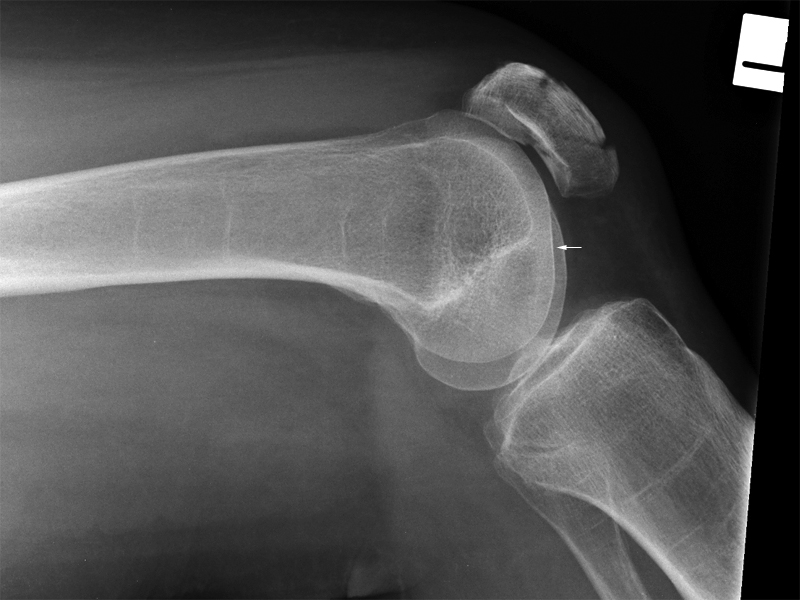
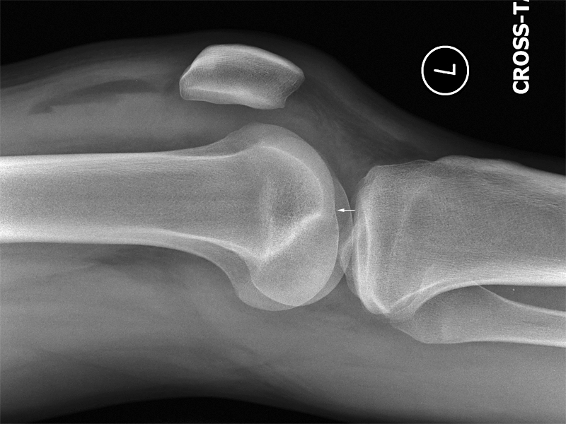
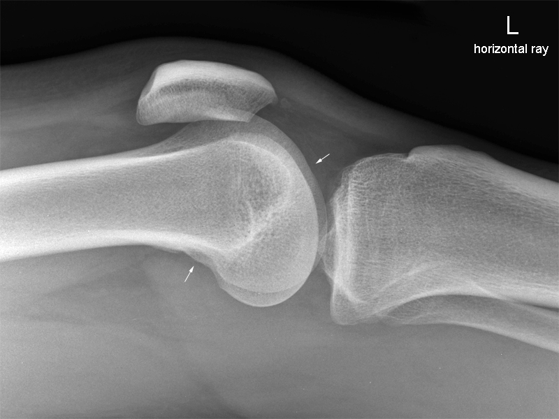
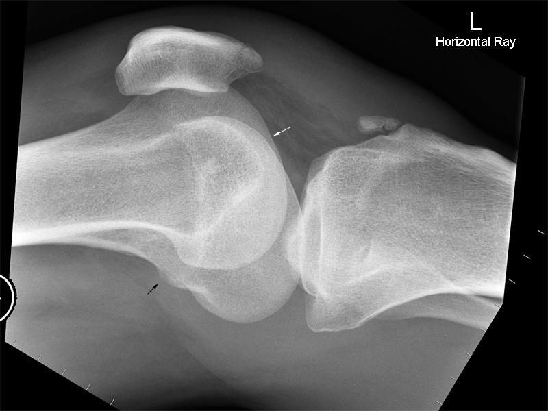
2. The lateral femoral notch The lateral condylopatellar sulcus(arrowed) , also known as the lateral femoral notch, distinguishes the lateral femoral condyle from the medial femoral condyle.
The lateral condylopatellar sulcus, also known as the lateral femoral notch, normally forms a shallow groove in the middle of the lateral femoral condyle. It represents the junction zone on the lateral femoral condyle where the tibiofemoral and patellofemoral radii of curvature meet. ... This appearance of the lateral sulcus also facilitates distinction between the lateral femoral condyle and the overlapping medial femoral condyle on the lateral projection.
<a class="external" href="http://radiology.rsna.org/content/219/3/800.full" rel="nofollow" target="_blank">Duke G. Pao, MD
The Lateral Femoral Notch Sign
June 2001 Radiology, 219, 800-801.
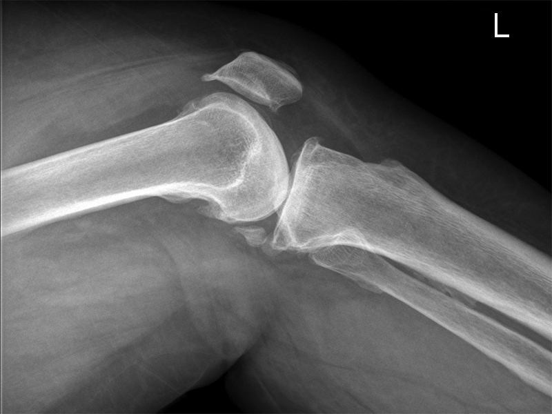
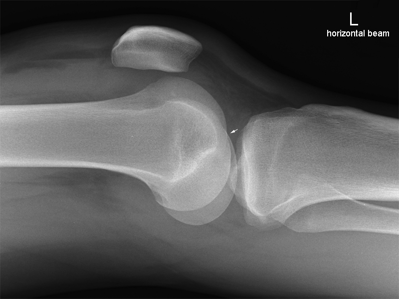
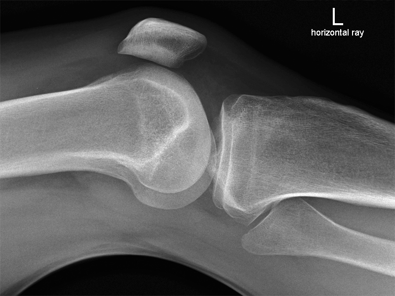
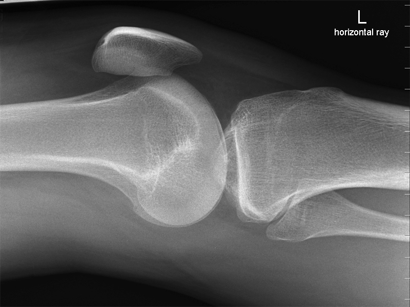
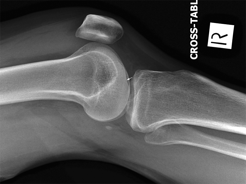
http://radiology.rsna.org/content/219/3/800.full</a>The repeat lateral knee image is shown left. The radiographer has reduced the degree of external rotation of the leg resulting in an improved lateral knee position. The lateral femoral notch is arrowed. Note that the fibula head is now largely superimposed over the tibial metaphysis.
3. The Adductor Tubercle (medial condyle)
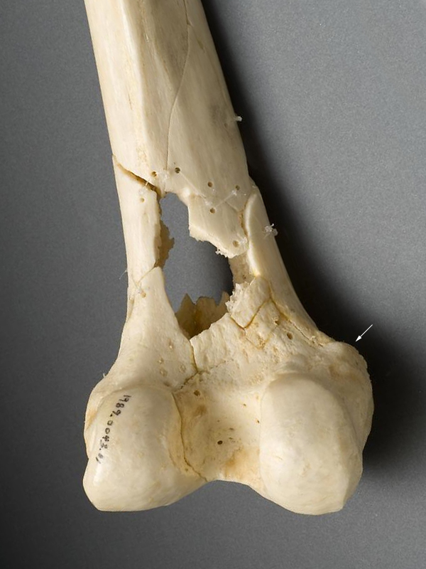
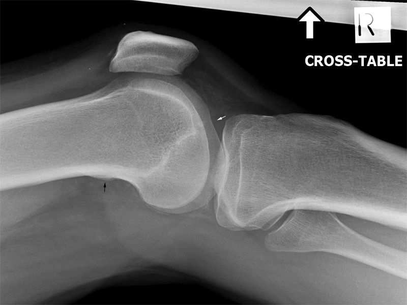
<a class="external" href="http://www.nlm.nih.gov/visibleproofs/media/detailed/ii_a_116a.jpg" rel="nofollow" target="_blank">adapted from http://www.nlm.nih.gov/visibleproofs/media/detailed/ii_a_116a.jpg</a>If you can imagine that you are looking at the back of your patient's left knee, this is what the distal femur would look like (may not be fractured like this though!). The smoothly surfaced adductor tubercle is on the medial side just proximal to the posterior aspect of the medial femoral condyle. This bony prominence can sometimes be utilised to differentiate the medial femoral condyle from the lateral femoral condyle when attempting to assess a malpositioned lateral knee image. The most reliable method for identifying the medial condyle is to locate the rounded bony tubercle known as the adductor tubercle.
[tubercle knee x-ray&source=bl&ots=t_ZLaTNpKi&sig=r-YzzoP-gS0gB8gPQONt0p24fp4&hl=en&ei=vLCDS66LPIqOkQWN3pCDAw&sa=X&oi=book_result&ct=result&resnum=1&ved=0CAUQ6AEwADgU#v=onepage&q=&f=false|Kathy McQuillen-MartensenRadiographic image analysis (2nd ed)Kathy McQuillen-MartensenElsevier Health Sciences, 2006, p319]
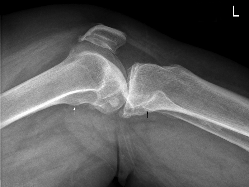
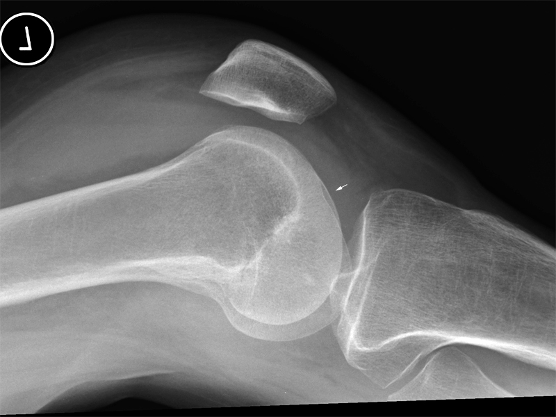
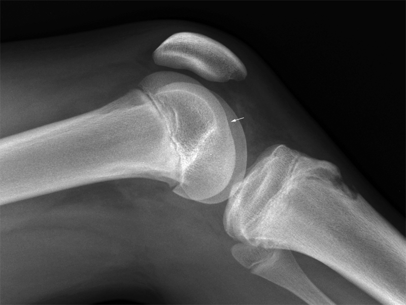
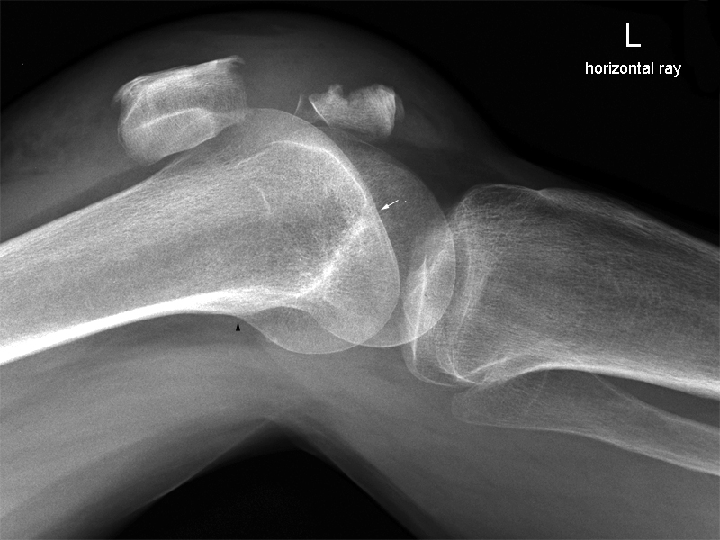
This lateral knee is malpositioned. The demonstration of the adductor tubercle (white arrow) allows differentiation of the medial femoral condyle from the lateral femoral condyle. The fibula head is excessively superimposed over the tibial metaphysis. These two observations suggest that the knee is too internally rotated.
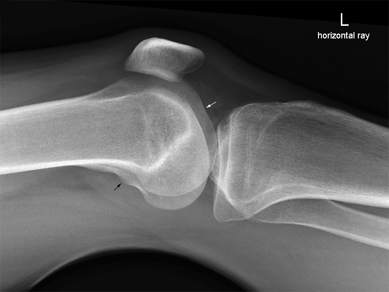
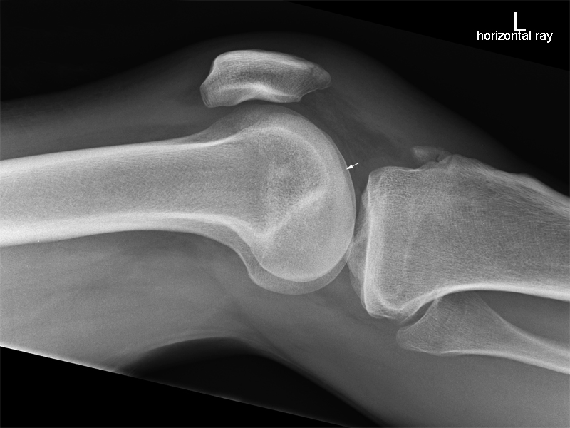
The radiographer repeated the lateral projection with the knee in a more externally rotated position (see below left).The repeat lateral knee shows good superimposition of the posterior aspects of the femoral condylar articular surfaces.
Note
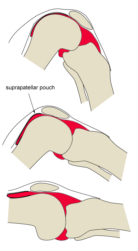
- improved demonstration of the suprapatellar pouch
- extensive degenerative disease of the knee joint and the patello-femoral joint
- fabella
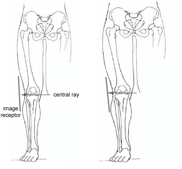
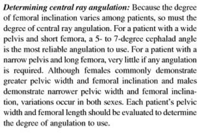
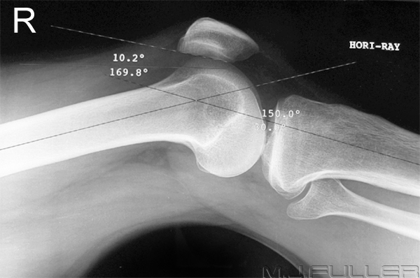
How much Tube Angulation is Required?
How much Knee Flexion is Required?
The Effect of Knee Flexion on the Patella and Suprapatellar Pouch
adapted from <a class="external" href="http://upload.wikimedia.org/wikipedia/commons/f/f3/Knee-unfolding-recess-diagram.svg" rel="nofollow" target="_blank">http://upload.wikimedia.org/wikipedia/commons/f/f3/Knee-unfolding-recess-diagram.svg</a>
Radiographic Technique
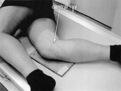
Horizontal Ray
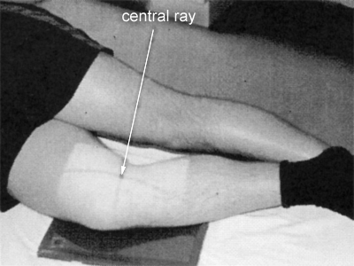
Rolled Lateral
Which Rolled Lateral Knee Radiography Technique is Superior?
The superior technique is the one that works for you. Research published in Synergy in 2006 (Symes, E. Lateral Knee Radiographs: Investigating the Techniques, Synergy, July 2006, p18) reported that 43% of the radiographers preferred technique 1 and 57 % preferred technique 2 (to the authors and my surprise!). The research also reported that radiographers did not always know why a lateral knee position failed (and presumably therefore did not know how to correct the malposition).
Case 1
Case 2
The Indicators
- The lateral femoral notch is demonstrated (white arrow)
- The adductor tubercle is not well demonstrated (although probably visible)
- The fibula head is not superimposed over the tibial metaphysis suggesting excessive external rotation of the knee.
For Horizontal Ray Lateral Technique
- To Correct Rotation Error: Rotate leg internally
- To Correct Angulation Error: More cephalic angulation
For Rolled Lateral technique
- To Correct Rotation Error: Rotate leg internally
- To Correct Angulation Error: More caudal Angulation
Note:
- Horizontal ray technique will always require opposite angulation correction to rolled technique because one technique is a medial-lateral beam and the other is lateral-medial beam
- These images are presented for the purpose of demonstration and are not necessarily worthy of repeating. The decision to repeat should be based on a balanced consideration of the costs vs the likely benefits to the patient
Discussion
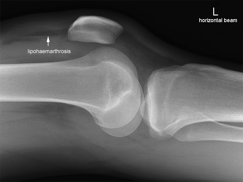
- There is a large knee effusion evident in Hoffa's fatpad and the suprapatellar pouch
- There is probably a lipohaemarthrosis
The Indicators
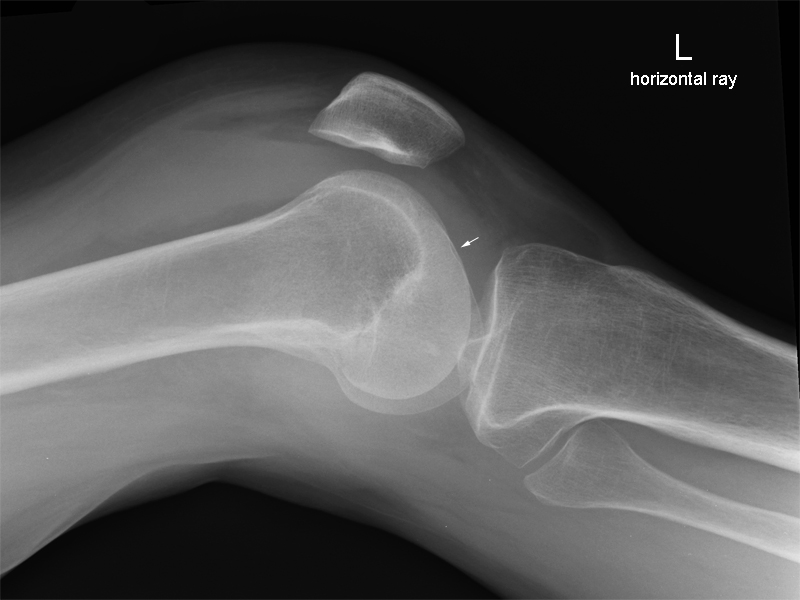
- The lateral femoral notch is demonstrated (white arrow)
- The adductor tubercle is not well demonstrated (although probably visible)
- The fibula position suggests too much external rotation
For Horizontal Ray Lateral Technique
- To Correct Rotation Error: Rotate leg internally
- To Correct Angulation Error: More cephalic angulation
For Rolled Lateral technique
- To Correct Rotation Error: Rotate leg internally
- To Correct Angulation Error: More caudal Angulation
Note
- Horizontal ray technique will always require opposite angulation correction to rolled technique because one technique is a medial-lateral beam and the other is lateral-medial beam
- These images are presented for the purpose of demonstration and are not necessarily worthy of repeating. The decision to repeat should be based on a balanced consideration of the costs vs the likely benefits to the patient
Discussion
- There is a large knee effusion evident in Hoffa's fatpad and the suprapatellar pouch
- There is a lipohaemarthrosis
- The lateral condylar sulcus is largely obscured by the overlying medial condylar articular cortcal bone
Case 3
The Indicators
- The lateral femoral notch is demonstrated (top white arrow)
- The adductor tubercle is demonstrated (bottom white arrow)
- The fibula head appears to be excessively overlapping the proximal tibial metaphysis
For Horizontal Ray Lateral Technique
- To Correct Rotation Error: Rotate leg externally
- To Correct Angulation Error: More cephalic angulation
For Rolled Lateral technique
- To Correct Rotation Error: Rotate leg externally
- To Correct Angulation Error: More caudal Angulation
Note:
- Horizontal ray technique will always require opposite angulation correction to rolled technique because one technique is a medial-lateral beam and the other is lateral-medial beam
- These images are presented for the purpose of demonstration and are not necessarily worthy of repeating. The decision to repeat should be based on a balanced consideration of the costs vs the likely benefits to the patient
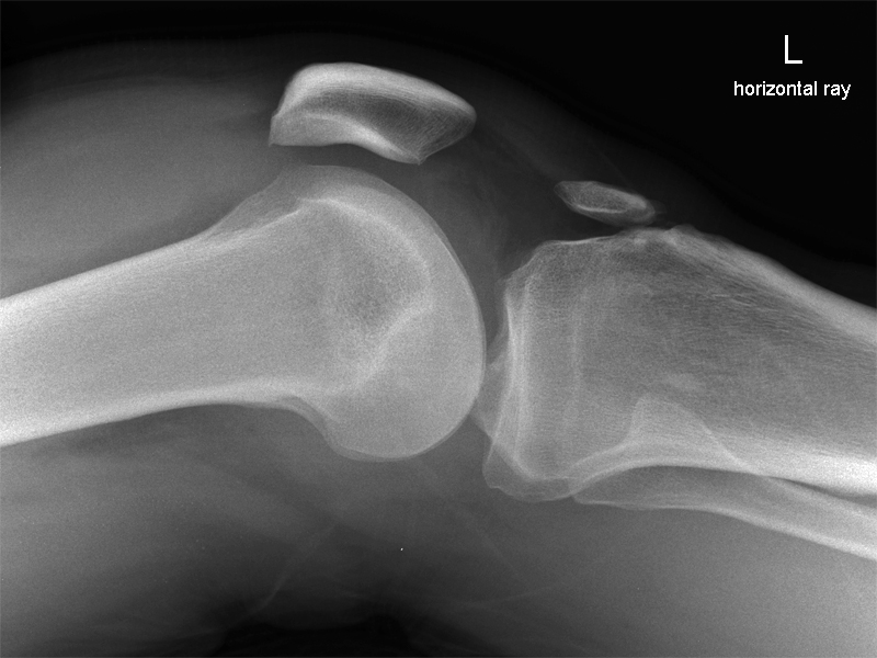
The Indicators
- The lateral femoral notch is not demonstrated
- The adductor tubercle is not demonstrated
- The fibula head is largely projected free of the proximal tibial metaphysis suggesting excessive external rotation of the knee.
For Horizontal Ray Lateral Technique
- To Correct Rotation Error: Rotate leg internally
- To Correct Angulation Error: More cephalic angulation
For Rolled Lateral technique
- To Correct Rotation Error: Rotate leg internally
- To Correct Angulation Error: More caudal Angulation
Note:Discussion
- Horizontal ray technique will always require opposite angulation correction to rolled technique because one technique is a medial-lateral beam and the other is lateral-medial beam
- These images are presented for the purpose of demonstration and are not necessarily worthy of repeating. The decision to repeat should be based on a balanced consideration of the costs vs the likely benefits to the patient
- This is a repeat of the lateral knee shown above. The radiographer has over-corrected the malposition error shown above.
- The radiographer has attempted to correct the rotation error only - the tube angle error is unchanged.
Case 4
The Indicators
- The lateral femoral notch is demonstrated (top white arrow)
- The adductor tubercle is demonstrated (bottom white arrow)
- The head of fibula is too superimposed over the proximal tibial metaphysis suggesting the need for further external rotation
For Horizontal Ray Lateral Technique
- To Correct Rotation Error: Rotate leg externally
- To Correct Angulation Error: More cephalic angulation
For Rolled Lateral technique
- To Correct Rotation Error: Rotate leg externally
- To Correct Angulation Error: More caudal Angulation
Note:
Discussion
- Horizontal ray technique will always require opposite angulation correction to rolled technique because one technique is a medial-lateral beam and the other is lateral-medial beam
- These images are presented for the purpose of demonstration and are not necessarily worthy of repeating. The decision to repeat should be based on a balanced consideration of the costs vs the likely benefits to the patient
- This lateral knee position would generally be considered within acceptable positioning limits
Case 5
The Indicators
- The lateral femoral notch is demonstrated (white arrow)
- The adductor tubercle is not demonstrated
- The fibula head position is normal
For Horizontal Ray Lateral Technique
- To Correct Rotation Error: no change required
- To Correct Angulation Error: More caudal angulation
For Rolled Lateral technique
- To Correct Rotation Error: no change required
- To Correct Angulation Error: More cephalic Angulation
Note:
- Horizontal ray technique will always require opposite angulation correction to rolled technique because one technique is a medial-lateral beam and the other is lateral-medial beam
- These images are presented for the purpose of demonstration and are not necessarily worthy of repeating. The decision to repeat should be based on a balanced consideration of the costs vs the likely benefits to the patient
Case 6
The Indicators
- The lateral femoral notch is demonstrated (white arrow)
- The adductor tubercle is not demonstrated
- The fibula head is largely unobscured by the proximal tibial metaphysis suggesting excessive external rotation of the knee
For Horizontal Ray Lateral Technique
- To Correct Rotation Error: Rotate leg internally
- To Correct Angulation Error: no change required
For Rolled Lateral technique
- To Correct Rotation Error: Rotate leg internally
- To Correct Angulation Error: no change required
Note:
Discussion
- Horizontal ray technique will always require opposite angulation correction to rolled technique because one technique is a medial-lateral beam and the other is lateral-medial beam
- These images are presented for the purpose of demonstration and are not necessarily worthy of repeating. The decision to repeat should be based on a balanced consideration of the costs vs the likely benefits to the patient
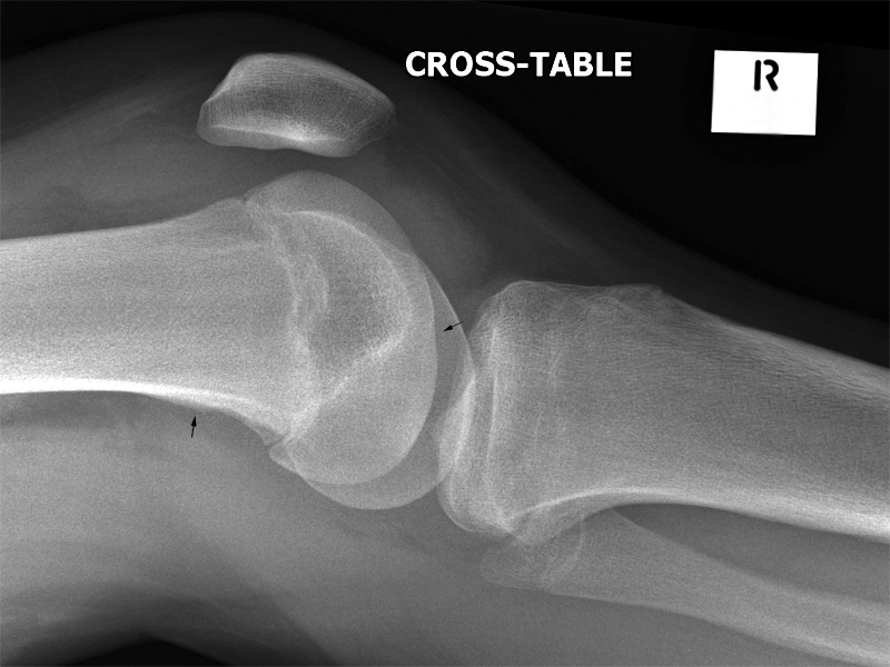
- this lateral knee position is acceptable and would not need repeating under normal circumstances.
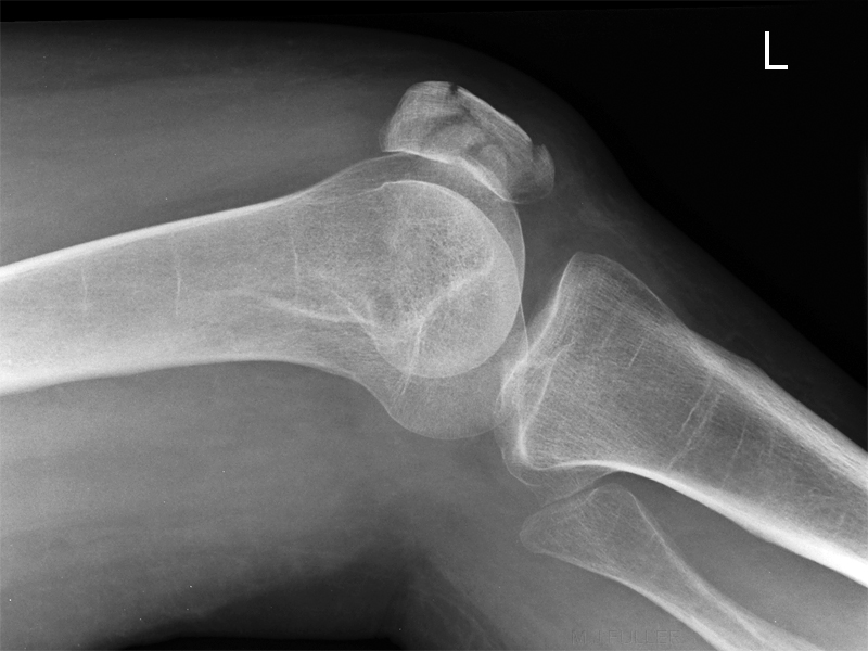
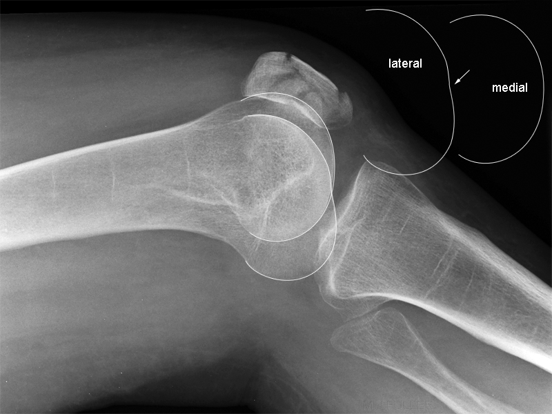
The Indicators
- The lateral femoral notch is demonstrated (white arrow)
- The adductor tubercle is demonstrated (black arrow)
- The fibula head is largely overlying the proximal tibial metaphysis suggesting excessive internal rotation positioning error.
For Horizontal Ray Lateral Technique
- To Correct Rotation Error: Rotate leg externally
- To Correct Angulation Error: More caudal angulation
For Rolled Lateral technique
- To Correct Rotation Error: Rotate leg externally
- To Correct Angulation Error: More cephalic Angulation
Note:
- Horizontal ray technique will always require opposite angulation correction to rolled technique because one technique is a medial-lateral beam and the other is lateral-medial beam
- These images are presented for the purpose of demonstration and are not necessarily worthy of repeating. The decision to repeat should be based on a balanced consideration of the costs vs the likely benefits to the patient
- patellar fracture noted
Case 7
The Indicators
- The lateral femoral notch is not demonstrated
- The adductor tubercle is not demonstrated
- The fibula head position is unremarkable
For Horizontal Ray Lateral Technique
- To Correct Rotation Error: no change required
- To Correct Angulation Error: ?
For Rolled Lateral technique
- To Correct Rotation Error: no change required
- To Correct Angulation Error: ?
Note
- Horizontal ray technique will always require opposite angulation correction to rolled technique because one technique is a medial-lateral beam and the other is lateral-medial beam
- These images are presented for the purpose of demonstration and are not necessarily worthy of repeating. The decision to repeat should be based on a balanced consideration of the costs vs the likely benefits to the patient
Comment
- You will sometimes see a lateral knee where all of the malposition indicators are absent
- knee joint effusion noted
- ? lipohaemarthrosis
Case 8
The Indicators
- The lateral femoral notch is demonstrated (white arrow)
- The adductor tubercle is demonstrated (black arrow)
- The fibula head position is unremarkable
For Horizontal Ray Lateral Technique
- To Correct Rotation Error: there is insignificant error
- To Correct Angulation Error: More cephalic angulation
For Rolled Lateral technique
- To Correct Rotation Error: there is insignificant error
- To Correct Angulation Error: More caudal angulation
Note
- Horizontal ray technique will always require opposite angulation correction to rolled technique because one technique is a medial-lateral beam and the other is lateral-medial beam
- These images are presented for the purpose of demonstration and are not necessarily worthy of repeating. The decision to repeat should be based on a balanced consideration of the costs vs the likely benefits to the patient
Case 9
The Indicators
- The lateral femoral notch is not well demonstrated (although probably visible)
- The adductor tubercle is not demonstrated
- The fibula head position is normal
For Horizontal Ray Lateral Technique
- To Correct Rotation Error: none
- To Correct Angulation Error: none
For Rolled Lateral technique
- To Correct Rotation Error: none
- To Correct Angulation Error: none
Note
- Horizontal ray technique will always require opposite angulation correction to rolled technique because one technique is a medial-lateral beam and the other is lateral-medial beam
- These images are presented for the purpose of demonstration and are not necessary worthy of repeating. The decision to repeat should be based on a balanced consideration of the costs vs the likely benefits to the patient
Case 10
The Indicators
- The lateral femoral notch is poorly demonstrated (top black arrow)
- The adductor tubercle is also poorly demonstrated (bottom black arrow)
For Horizontal Ray Lateral Technique
- To Correct Rotation Error: insignificant error
- To Correct Angulation Error: increased caudal angulation
For Rolled Lateral technique
- To Correct Rotation Error: insignificant error
- To Correct Angulation Error: decreased caudal angulation
Note
- Horizontal ray technique will always require opposite angulation correction to rolled technique because one technique is a medial-lateral beam and the other is lateral-medial beam
- These images are presented for the purpose of demonstration and are not necessarily worthy of repeating. The decision to repeat should be based on a balanced consideration of the costs vs the likely benefits to the patient
Discussion
- knee joint effusion noted
Case 11
The Indicators
- The lateral femoral notch is not demonstrated
- The adductor tubercle is not well demonstrated
- The fibula head position is unremarkable
For Horizontal Ray Lateral Technique
- To Correct Rotation Error: none
- To Correct Angulation Error: none
For Rolled Lateral technique
- To Correct Rotation Error: none
- To Correct Angulation Error: none
Note
- Horizontal ray technique will always require opposite angulation correction to rolled technique because one technique is a medial-lateral beam and the other is lateral-medial beam
- These images are presented for the purpose of demonstration and are not necessary worthy of repeating. The decision to repeat should be based on a balanced consideration of the costs vs the likely benefits to the patient
Discussion
- large knee joint effusion noted
Case 12
The Indicators
- The lateral femoral notch is demonstrated (white arrow)
- The adductor tubercle is not demonstrated
- The fibula head position is normal
For Horizontal Ray Lateral Technique
- To Correct Rotation Error: none
- To Correct Angulation Error: none
For Rolled Lateral technique
- To Correct Rotation Error: none
- To Correct Angulation Error: none
Note
- Horizontal ray technique will always require opposite angulation correction to rolled technique because one technique is a medial-lateral beam and the other is lateral-medial beam
- These images are presented for the purpose of demonstration and are not necessarily worthy of repeating. The decision to repeat should be based on a balanced consideration of the costs vs the likely benefits to the patient
Discussion
- knee joint effusion noted
Case 13
The Indicators
- The lateral femoral notch is poorly demonstrated (white arrow)
- The adductor tubercle is also somewhat poorly demonstrated (black arrow)
- The fibula head position is normal
For Horizontal Ray Lateral Technique
- To Correct Rotation Error: none
- To Correct Angulation Error: More caudal angulation
For Rolled Lateral technique
- To Correct Rotation Error: none
- To Correct Angulation Error: More cephalic Angulation
Note
- Horizontal ray technique will always require opposite angulation correction to rolled technique because one technique is a medial-lateral beam and the other is lateral-medial beam
- These images are presented for the purpose of demonstration and are not necessarily worthy of repeating. The decision to repeat should be based on a balanced consideration of the costs vs the likely benefits to the patient
Discussion
- Fractured patella noted
Case 14
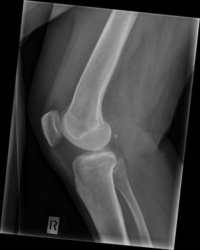
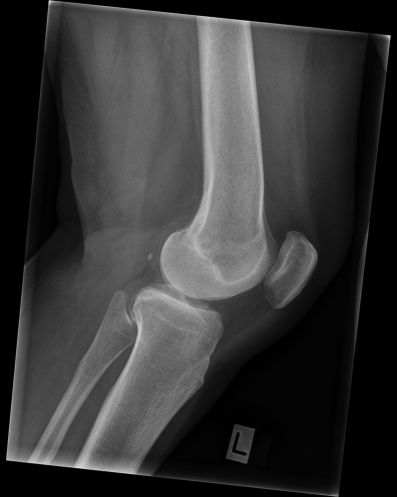
This 73 year old lady presented with right knee pain and a history of gout. She was referred for radiography of both knees. You can be confident that you have mastered lateral knee radiography when you are producing good quality images... you can be very confident when you produce bilateral knee images that look like mirror images!
Comment
Lateral knee radiography is one of the more difficult radiographic positioning challenges (perhaps surpassed only by the lateral elbow). Persistence and practice will yield results. If you have any technique tips, you can post them as a thread on this page.
... back to the Applied Radiography home page
... back to the Wikiradiography home page
