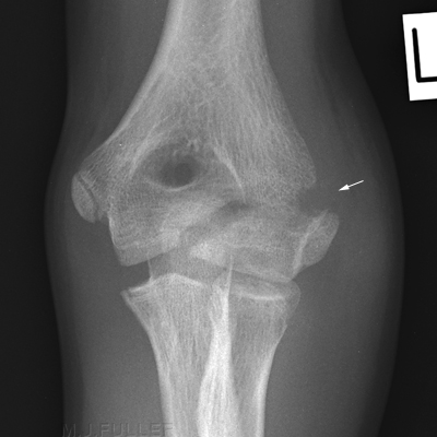
Lateral Condylar Elbow Fractures
Fractures of the lateral humeral condyle are the second most common fracture of the paediatric elbow (John Harris et al The Radiology of Emergency Medicine, 3rd Ed, Williams and Wilkins, 1993, p 352). This page examines the radiographic demonstration of lateral condyle fractures of the elbow.
The Salter-Harris Classification
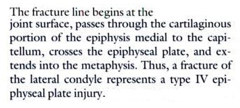
Classification of Lateral Condyle FracturesSalter notes that the lateral condyle fractures of the distal humerus are Salter Harris 4 fractures..
<a class="external" href="http://books.google.com.au/books?id=oa6fDFuX-I8C&pg=PA523&lpg=PA523&dq=elbow+condyle+vs+epicondyle&source=bl&ots=lhiHITotg0&sig=YDhHTE7q_t_nQN0pcYGHEr0kFqo&hl=en&ei=wSBPSrS3Loj8sQPOvYWrDQ&sa=X&oi=book_result&ct=result&resnum=7" rel="nofollow" target="_blank">(Robert. Bruce. Salter ,</a>
<a class="external" href="http://books.google.com.au/books?id=oa6fDFuX-I8C&pg=PA523&lpg=PA523&dq=elbow+condyle+vs+epicondyle&source=bl&ots=lhiHITotg0&sig=YDhHTE7q_t_nQN0pcYGHEr0kFqo&hl=en&ei=wSBPSrS3Loj8sQPOvYWrDQ&sa=X&oi=book_result&ct=result&resnum=7" rel="nofollow" target="_blank">Textbook. of Disorders and Injuries of the Musculoskeletal System. 3rd Ed, 1999, p523)</a>
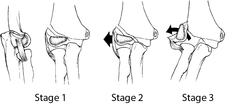
Classification of Lateral Condylar Elbow Fractures
<a class="external" href="http://www.orthospot.com.au/papers.orthospot.com.au/fracupl_files/frame.htm" rel="nofollow" target="_blank">http://www.orthospot.com.au/papers.orthospot.com.au/fracupl_files/frame.htm</a>This classification system is largely self-explanatory.
Case 1
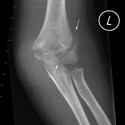
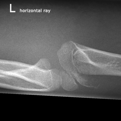
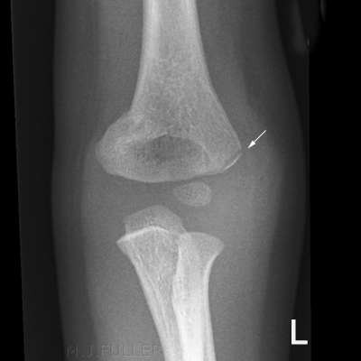
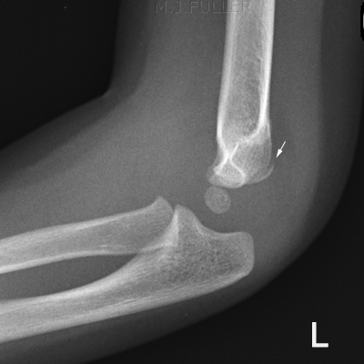
This 6 year old girl presented to the Emergency Department after an unwitnessed fall. She was refusing to use her left arm and her left elbow was painful and swollen. She was referred for radiography of her left elbow.
Case 2
This 2 1/2 year old girl presented to the Emergency Department after an unwitnessed fall. She was refusing to use her left arm and her left elbow was painful and swollen. She was referred for radiography of her left elbow.
Case 3
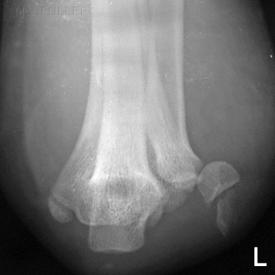
The details of this case are unknown
Comment
This case demonstrates the potential value of the axial view of the elbow in cases of lateral condyle facture.
Case 4
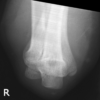
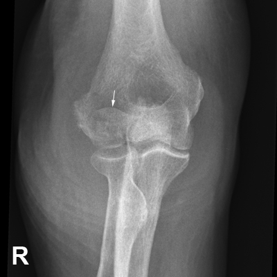
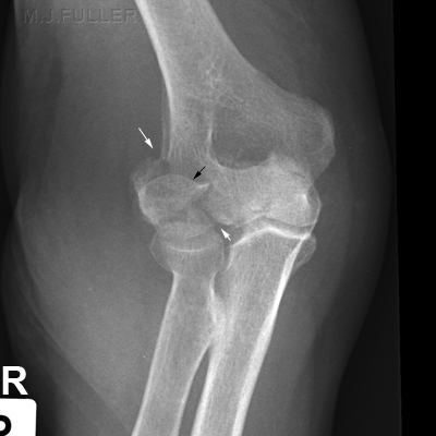
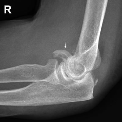
This 81 year old lady presented to the Emergency Department after falling onto her right side. Her right elbow was painfull and swollen. She was referred for radiography of her right elbow.
Case 5
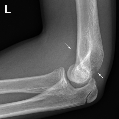
This 14 year old male presented to the Emergency Department with a painful left elbow. The mechanism of injury is unknown. He was referred for radiography of his left elbow.
...back to the Wikiradiography home page
...back to the Applied Radiography home page
