Introduction With the introduction of digital radiography, new possibilities have opened up that have not existed before in the world of developer and fixer radiography. One of these new possibilities is the potential to extract two images from the one exposure. How valid is this technique when applied to patients who are referred for chest and thoracic spine radiography?
Case Study 1
Patient History
This elderly patient presented to the Emergency Department with spontaneous onset of chest and thoracic spine pain. The patient had a history of COAD and osteoporosis.
Technique
The radiographer has performed an erect PA and lateral chest X-ray examination using the AEC. The lateral chest image is shown below.
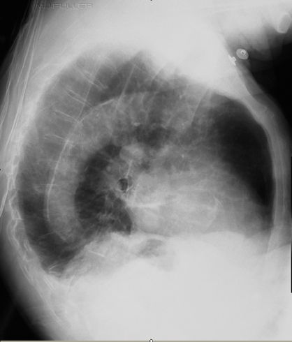 | When using digital radiography, it is tempting at this stage for the radiographer to: - apply post-processing collimation to this image;
- apply a lateral thoracic spine algorithm;
- and re-save the image as a lateral thoracic spine view.
This approach will save the patient an additional radiation dose and will prevent the patient from being subject to the unnecessary rigors of undertaking an additional lateral thoracic spine view. These laudable objectives must be offset against the fact that the lateral thoracic spine and lateral chest exposure techniques are diametrically opposed. The lateral chest radiography exposure technique uses a small exposure time to limit movement unsharpness of soft tissue structures. The lateral thoracic spine exposure technique uses a long exposure time technique to blur the soft tissues of the thorax.
Do you think the radiographer has demonstrated the lateral thoracic spine anatomy adequately on this lateral chest image?
The radiographer proceeded to undertake a lateral thoracic spine radiographic examination.
|
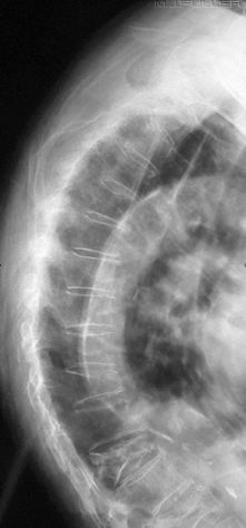
| A breathing lateral thoracic spine exposure technique was employed to good effect. The thoracic and upper lumbar vertebral bodies are clearly visualised. There is evidence of osteoporosis (pencilling of the vertebral bodies). There are several vertebral body wedge fractures with severe wedging of L1.
Compare this image with the thoracic vertebrae demonstrated on the lateral chest image above |
Case Study 2
Patient History
This 80 year old lady presented to the Emergency Department with spontaneous onset of thoracic spine pain. The patient had a history of breast cancer. She was referred for chest radiography. The clinical question was whether her symptoms were related to chest pathology or thoracic vertebral body crush fracture(s).
Technique
The radiographer has performed an erect PA and lateral X-ray examination using the AEC. A Philips Digital Diagnost release I was utilised to produce these images.
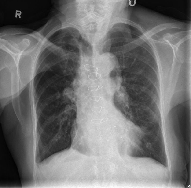 | The PA chest image demonstrates sternal sutures and 3 metal clips relating to previous cardiac surgery. There is an inferior accessory lung fissure on the right. There is asymmetry of lung density (darker on the right) of unknown aetiology (? technical rather than pathological). |
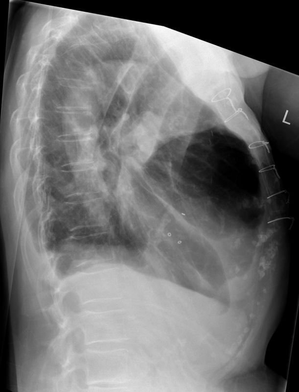 | The lateral chest image demonstrates a possible small interlobar effiusion (? right sided). There is some loss of clarity of the left hemidiaphragm posteriorly of uncertain aetiology.
The thoracic vertebral bodies show penciling consistent with osteopenia. This is likely to be associated with osteoporosis. |
Discussion
It is noteworthy that the clinical question included the possibility of the patient's pain being related to thoracic vertebral body crush fracture(s). Despite this provisional diagnosis offered by the referring doctor, thoracic spine radiography was not requested. The imaging was reviewed by the referring doctor and the patient was referred for thoracic spine radiography.
- Is thoracic spine radiography warranted when the thoracic spine has been demonstrated on the chest radiography?
- Should the referring doctor have request a CXR and thoracic spine radiography in the first instance?
- Should the radiographer have performed thoracic spine radiography at the same time as the chest radiograph was performed? (either within the radiographer's scope of practice or by communicating their concerns to the referring doctor)
|
|
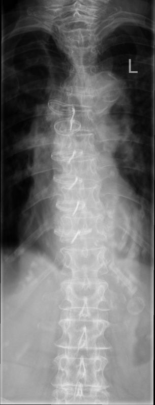 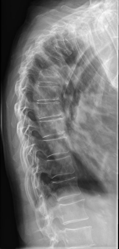 | The radiographer noted that the patient was complaining of high thoracic spine pain "between the shoulder blades". This was the least well demonstrated part of the thoracic spine on the lateral chest image. It is also noteworthy that there appeared to be loss of height of some of the upper thoracic vertebral bodies demonstrated on the lateral chest image.
Thoracic spine radiography was performed. The radiography was performed on a Philips Digital Diagnost release II using a fixed kVp, mA and exposure time (manual exposure technique). The exposure time for both images is 1.0 second.
The AP thoracic spine image demonstrates blurring of the soft tissue structures associated with the breathing technique. The thoracic spine pedicles are largely well demonstrated- this could be important in a patient with thoracic spine pain and a known history of breast cancer- the pedicles are a common site of secondary metastatic deposits. A button artifact is demonstrated to the left of L1.
The radiographer employed a breathing technique for the lateral thoracic spine projection to good effect. The lateral thoracic spine image demonstrates loss of height of several of the upper thoracic vertebral bodies. These could reflect recent vertebral body crush fractures and may account for the patient's symptoms.
On balance, the radiographer should have considered the possibility of performing dedicated thoracic spine radiography at the time of the chest radiography given the following
- the referring doctor had stated on the referral that thoracic spine crush fractures were one of the provisional diagnoses
- The patient was complaining of pain "between the shoulder blades"
- There was evidence of vertebral body crush fractures on the lateral chest X-ray image.
- Secondary tumour deposits in thoracic pedicles from the known breast cancer would probably have been missed on the PA chest image
|
Discussion It is clear that a breathing exposure technique can have a significant beneficial effect on the quality of thoracic spine imaging. This is not to suggest that this approach should be applied universally when the thoracic spine has been well demonstrated on chest radiography. Indeed, there may be occasions where, on balance, "digital double dipping" is the correct choice. This is a judgement that can and should be made by the radiographer. You should have a low threshold for discussing the options with senior radiography staff, radiologist and/or the referring doctor where the choice is not clear.
Good radiography is that which is the best yield/risk for that patient,
at that time, and for their likely conditions.
....back to the applied radiography home page here

