Lateral Chest Paravertebral Gutter Positioning Technique
What constitutes a lateral chest radiographically? This page considers paravertebral gutter technique as a means of producing a lateral chest position. Irrespective of what you consider a good lateral chest position to be, this technique may have some utility. This page describes a practical approach to lateral chest radiography technique.
The True Lateral Chest
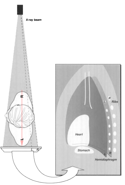
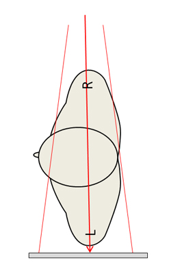
adapted from <a class="external" href="http://books.google.com.au/books?id=FBYrSSys_LsC&printsec=frontcover#PPA14,M1" rel="nofollow" target="_blank">Jannette Collins, Eric J. Stern</a><a class="external" href="http://books.google.com.au/books?id=FBYrSSys_LsC&printsec=frontcover#PPA14,M1" rel="nofollow" target="_blank">Chest Radiology: The Essentials Edition: 2, 2007</a>This graphic illustrates the true lateral chest position. The problem with this position is how to achieve it consistently.
The Paravertebral Gutters
The Paravertebral Gutter Technique
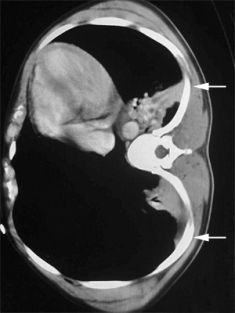
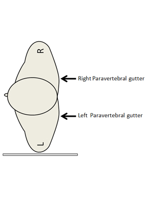
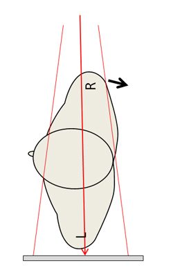
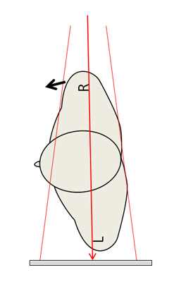
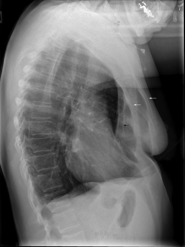
The paravertebral gutter technique for lateral chest radiography is one of those techniques that is very easy to execute but hard to explain. The general principle is that if you line up the skin adjacent to the paravertebral gutter with the divergent beam (light beam and X-ray beam), in doing so, you are also overlapping the posterior ribs. Position the patient in the true left lateral position with arms folded over his/her head. Rotate the patient's right side forward (anteriorly) until the light from the LBD just skims the left paravertebral gutter (i.e. the left paravertebral gutter is just out of the shadow of the right paravertebral gutter). In this position the posterior ribs are perfectly, or near-perfectly, superimposed.
Advantages and Disadvantages of the Paravertebral Gutter Technique/Position
Advantages Disadvantages
- Highly Reproducible
- Not a true lateral
- Can result in an oblique view of the sternum (trauma consideration)
- Does not work in patients who don't have visible paravertebral gutter surface anatomy (scoliosis, emphysema, obese patients)
- May superimpose posterior costophrenic angles
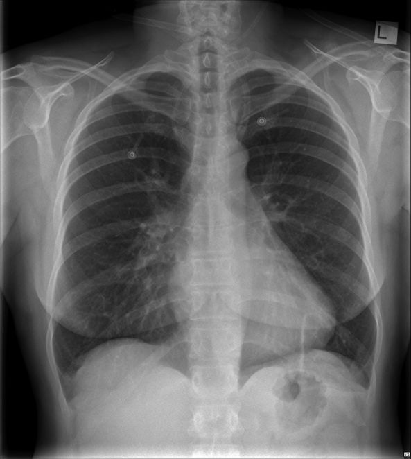
Case 1
Discussion
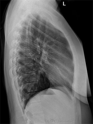
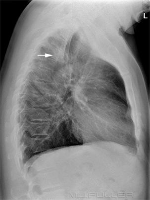
Superimposition of the posterior ribs on a lateral chest image is considered by some radiographers as a mark of a good lateral chest X-ray position. Conversely, it is considered a hallmark of bad technique in other institutions. In particular, some paediatric departments require separation of the posterior ribs on the lateral image to assist in separating the posterior costophrenic angles. It must be frustrating for a radiologist to identify a basal lung pathology on a lateral chest image, but not know which side of the chest is involved.
The use of the paravertebral gutter technique to superimpose the posterior ribs allows radiographers to achieve consistent and repeatable lateral chest positioning. This raises the question of whether the ability to reproduce the exact same comparable lateral chest position is of greater utility than producing images that attempt to meet the goal of a true lateral chest position (with unproven diagnostic benefit). Equally, using the paravertebral gutter technique may simply reflect tidymindedness on the part of the radiographer that is of no real benefit to the patient.
... back to the Applied Radiography home page
