Knee
Jump to navigation
Jump to search
Knee Surface Anatomy | Other pages of interest |
Knee Movements
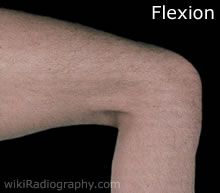
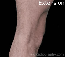
| | The knee is a hinge joint and can perform both flexion and extension. Movement is prevented in other directions as it is supported by a series of strong ligaments, menisci and the muscles which traverse the joint. |
Knee Anterior Surface
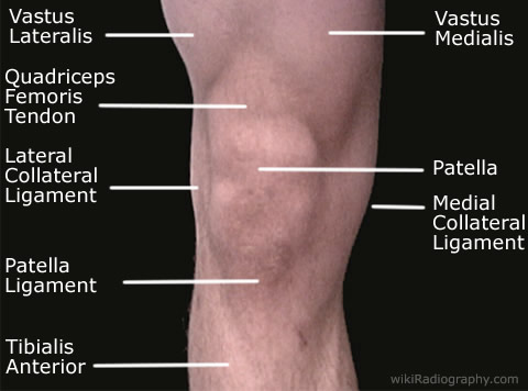
| | The vastus lateralis is the largest of the four muscles in the hamstring muscle group, its main function is to provide extension of the knee. The vastus medialis is also part of the quadriceps group and the location of the orgin and insertion of the muscle show that it also provides extension of the knee. The lateral collateral ligament protects the lateral side from an inside bending (varus force).The medial collateral ligament protects the knee from being bent by a stress (valgus force) applied to the lateral side of the knee. The patelllar ligament which is often called the patellar tendon because there is no definite separation between the quadriceps tendon (which surrounds the patella) and the area connecting the patella to the tibia. This very strong ligament helps give the patella its mechanical leverage. The patella is a thick, triangular bone which articulates with the femur and covers and protects the front of the knee joint. It is the largest sesamoid bone in the human body. The tibialis anterior is a muscle that runs the length of the tibia. It acts to dorsiflex the foot. |
Knee Posterior Surface
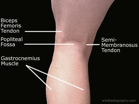
| | The gastrocnemius muscle is located in the superficial posterior compartment of the leg. It is involved with plantar flexion of the foot. The popliteal fossa is a the shallow depression located at the back of the knee-joint.The biceps femoris is a muscle of the posterior thigh, its tendon can be seen as it crosses over the posterior aspect of the knee joint. Its is involved in flexion of the knee. The semimembranosus is also a muscle in the posterior region of the thigh. Due to its origin and insertion it is involved in both flexion of the knee and extension at the hip. Its tendon can be seen as it crosses over the posterior aspect of the knee joint. |
Go Back to Surface Anatomy Index


