Is the Screw in the Joint?

From wikiRadiography
Revision as of 17:01, 11 November 2020 by Travis (talk | contribs) (Created page with "<div class="WPC-editableContent"><div align="center"><font size="4"><font color="#ff0000"><b><br/></b></font></font></div><font size="4"><b>Introduction</b></font><br/><blockq...")
(diff) ← Older revision | Latest revision (diff) | Newer revision → (diff)
Jump to navigation Jump to search

Introduction

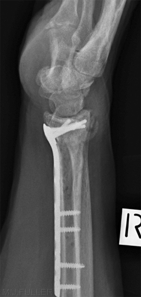
When an orthopaedic surgeon applies plates and screws to a wrist fracture, it is important that the screws do not penetrate into the wrist joint. Standard radiographic views of the wrist often project the tips of the screws into the wrist joint. This page considers radiographic techniques that will demonstrate the true position of these screws in relation to the wrist joint.


Radiography

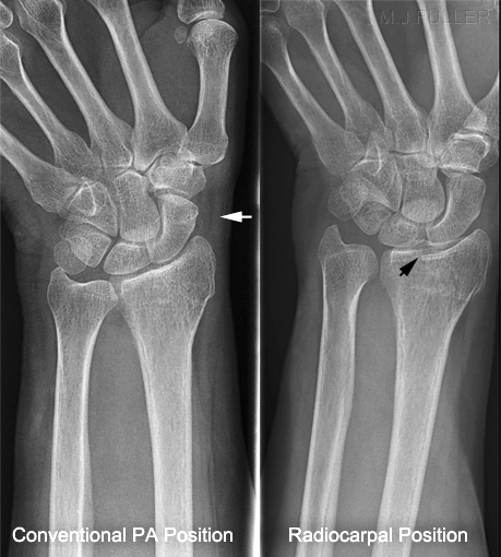
When standard PA and lateral views of the wrist are performed, the radiocarpal joint is not imaged in profile

radial inclinationradial inclinationlateral Wrist
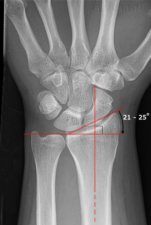
Radial inclination
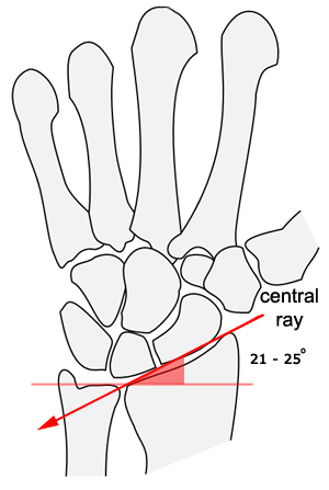
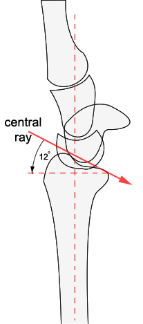
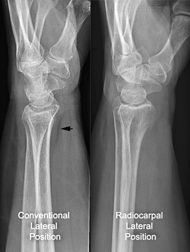
To demonstrate the radiocarpal joint, the lateral view of the wrist requires a beam angulation (or positioning equivalent) to match the patient's radial inclination.
To demonstrate the radiocarpal joint on a lateral wrist projection, the central ray should be angled as shown above. Note the demonstration of the joint between radius and lunate


palmar tiltpalmar tiltPA wrist
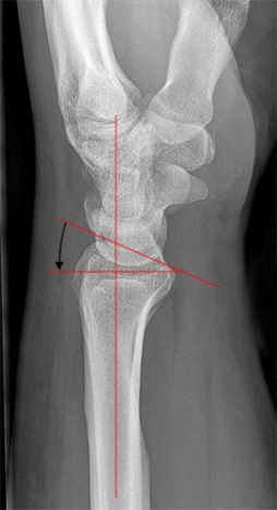
Palmar Tilt
The PA view would require beam angulation (or equivalent positioning ) to match the patient's palmar tilt.
The palmar tilt is variable, but will average about 12 degrees in an adult.The demonstration of the radiocarpal joint is not perfect, but will be an improvement on a conventional PA wrist position.


lateral wristlateral Wrist
lateral wristlateral wrist
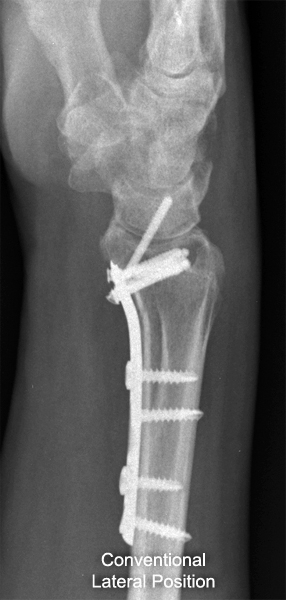
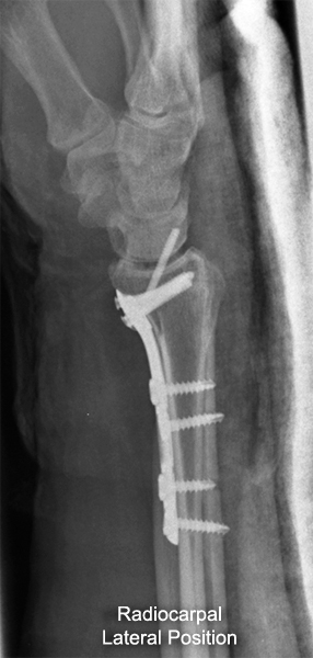
The conventional lateral wrist position requires no explanation. Note that the radiolunate joint is not well demonstratedThe modified radiocarpal lateral position requires the patient's hand to be raised and rested on a positioning sponge. The hand is raised to profile the distal radial joint surface. The distal inclination will vary from patient to patient (and may be altered post surgery) but will normally be in the range of 20 - 25 degrees.


The Radial Styloid Screw Trap

Radiocarpal View
lateral wristmodified lateral wrist
Radiocarpal View
PA wristmodified PA wrist
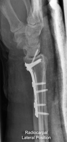
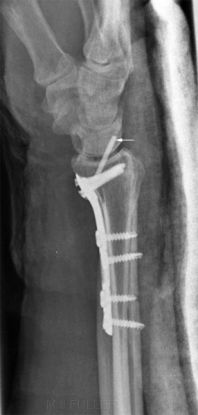
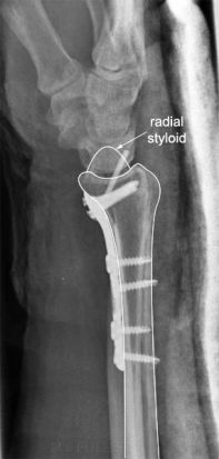
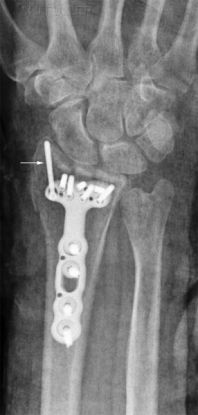
Even when a modified lateral wrist position is employed to demonstrate the radiocarpal joint, a single screw can appear to be within the wrist joint. This is commonly the longer radial styloid screw which is usually not in the joint. The reason that the radiocarpal view does not demonstrate the true tip position of this screw is that the distal radius is concave in both planes- you can't profile all of the joint in a single image.

Note: this screw is possibly protruding dorsally but still does not breach the articular surface of the radius.
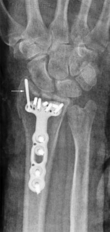
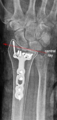
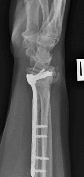
The PA view demonstrates the radial styloid screw to be external to the wrist joint.

The radiocarpal PA view of the wrist is less likely to provide an improved impression of the screw tip position than the lateral radiocarpal view because the palmar tilt (approx 12 degrees) is a smaller angle than the radial inclination (approx 22 degrees)


Case1
PA wristlateral wristlateral wrist
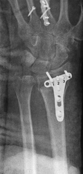
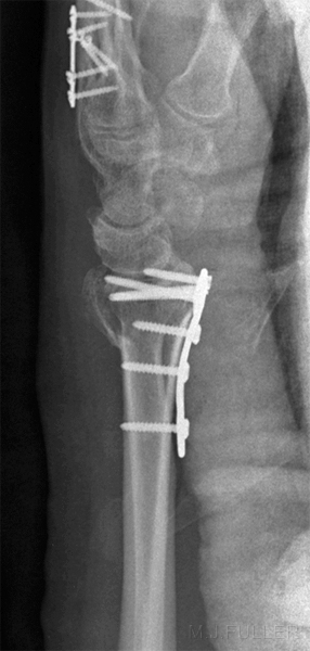
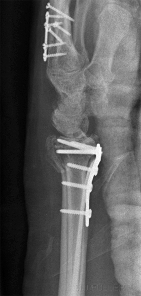
This is a conventional PA wrist image taken with the patient's wrist in a plaster cast. Screw 1 appears to be in contact with the patient's lunate.This is a conventional lateral wrist in plaster. Two of the screws appear to be partly within the wrist joint. Screw 4 is known not to be in the joint from the PA view.The radiocarpal lateral view suggests that screws 1 and 4 are close to the radial joint surface but do not protrude into the wrist joint

Comment

On the basis of the conventional views of the wrist, screw 1 appears to be within the radiocarpal joint. The modified radiocarpal lateral view image suggests that screw 1 is less of a threat to the radial articular cartilage than would be suggested by the conventional wrist positioning radiography.


Case 2
LATERAL WRISTLATERAL WRIST
This is a conventional lateral wrist image. One of the screws appears to be in contact with the patient's lunate.This is a radiocarpal modified lateral wrist position. The screw in question is demonstrated to be in less threatening position.


Case 3
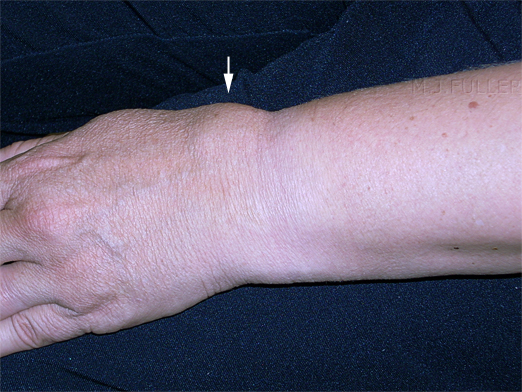
wrist fractureThis lady presented to the Emergency Department after a fall and was referred for wrist radiography. The images below show her wrist after the bandage was removed. She was in considerable pain and the focal swelling over the dorsum of her wrist suggested that she may have sustained a fractured radius.


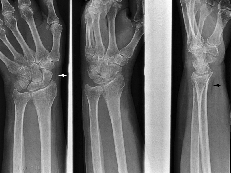
Routine wrist views were performed as shown below


wrist fracture
This series demonstrated no obvious fracture... although the distal radius is suspect looking. The scaphoid fat pad (white arrow) and the pronator fat pad (black arrow) were normal.


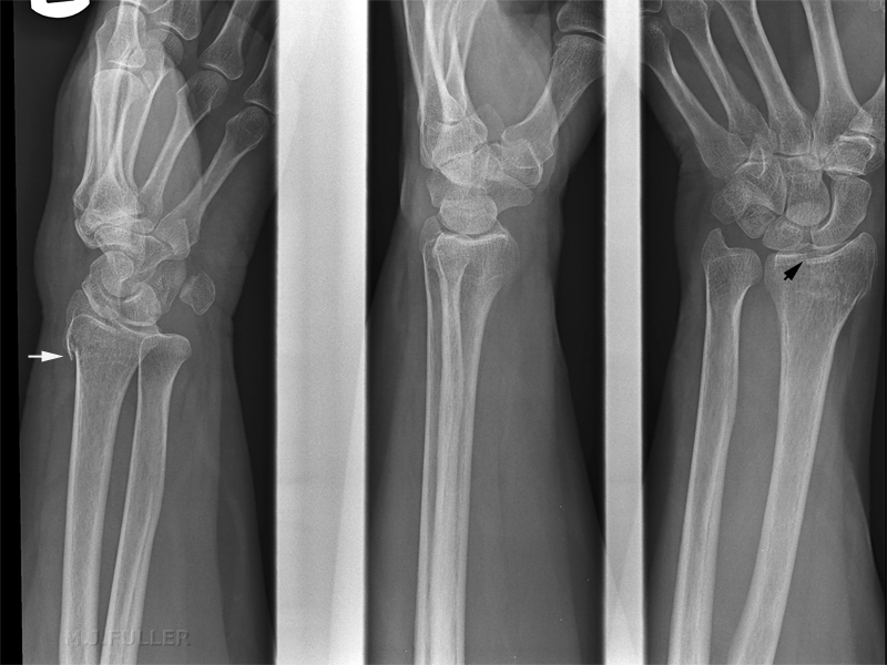
The radiographer was convinced clinically that the patient had sustained a fracture and considered whether the soft tissue signs were misleading (as there are known to be on occasions). Supplementary views were performed as shown below.

wrist fracture
The views performed are (from left to right)
  1. a reverse oblique wrist
  2. modified lateral wrist to profile articular surface of radius (radiocarpal view)
  3. modified pa wrist to profile the articular surface of radius (radiocarpal view)
The reverse oblique image demonstrates a fractured distal radius (white arrow). The fracture and step in the articular surface of the radius is also demonstrated on the modified PA image (black arrow).


radiocarpal PA wristradiocarpal lateral wrist
For the purposes of comparison, the two PA images are shown together above. Note that the step in the radial joint surface is clearly demonstrated in the radiocarpal view but hidden in the conventional PA wrist image.The two lateral views are difficult to compare because of the under-rotation of the radiocarpal view.


Comment

Orthopaedic surgeons often perform radiocarpal views of the wrist following ORIF as the final image intensifier images. This raises the question of whether we should be routinely performing the same modified wrist views when these patients are referred for follow-up imaging. The joint of interest in these cases is the radiocarpal joint- the articulation between the lunate/scaphoid and the distal radius articular surface- should we not be imaging specifically for this joint?

The radiocarpal views (both PA and lateral) have been found to be valuable supplementary views in patients with subtle distal radius fractures.

... back to the Wikiradiography home page
... back to the Applied Radiography page