Incisional Hernia
This patient presented to the Emergency Department with acute abdominal pain. Following a clinical examination, he was referred for an acute abdominal X-ray examination. The referring doctor provided the following clinical information “?Incisional hernia, ?incarcerated”.
Patient History
The patient has a history of previous abdominal surgery. This is an important part of the patient’s history. Adhesions from previous abdominal surgery are the most common cause of small bowel obstruction in western society. (1) In this case, the history of previous abdominal surgery is important for another reason- the patient has a tender lump on his anterior abdominal wall, raising the possibility of an incisional hernia.
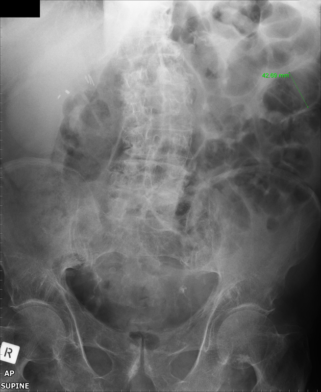
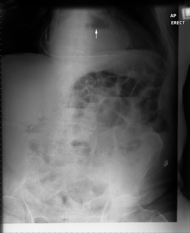
The patient had presented to the Emergency Department with acute abdominal pain 7 months earlier. The erect and supine abdominal images from this presentation are shown below
There are multiple loops of gas-filled small bowel predominantly in the left upper quadrant. One of these loops has a measured diameter (uncalibrated) of 43mm suggesting small bowel obstruction (SBO). Normal small bowel can have a diameter of up to 30mm. There is a paucity of gas in the large bowel which supports the diagnosis of SBO. Surgical staples are noted in the right upper quadrant. Lumbar degenerative disease and scoliosis concave to the right are noted. There is calcification of the abdominal aorta which is not clearly aneurysmal.
The erect abdominal image demonstrates a few fluid levels in a step-ladder configuration. There are pacemaker leads and sternal sutures demonstrated. A fluid level behind the heart shadow (arrowed) is likely to be contained within a hiatus hernia. This examination suggests SBO, which could be caused by incarceration of small bowel loops within an incisional hernia, from adhesions from previous surgery, or from some other cause.
Current Imaging
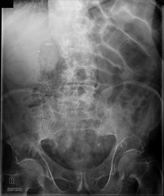
The images from the current presentation are shown below
The supine abdominal image shows similarities with the image from the earlier presentation. The diagnosis of SBO is more convincing with several loops of distended small bowel.
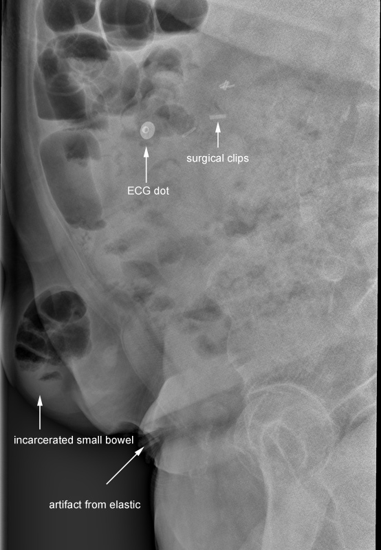
This image clearly demonstrates the incarcerated small bowel. Clinical correlation suggested that this incarcerated small bowel was within an incisional hernia. This image provides a confident diagnosis for the referring doctor/surgeon and may save the patient from undergoing further imaging.
Abdominal Hernias
Abdominal hernias are common abdominal pathologies. Abdominal hernias can be internal, external or diaphragmatic. Internal hernias are those in which there is an abnormal protrusion of the abdominal viscera into a normal or abnormal abdominal cavity, potential space or compartment. External hernias involve protrusion of the abdominal contents through a defect in the abdominal wall. Types of abdominal wall hernias include umbilical, spigelian, incisional, inguinal and femoral (2). An incisional hernia is an abnormal protrusion of abdominal contents through a surgical defect in the abdominal wall.
Radiographic Technique
The tangential view of an incisional hernia can be performed in the lateral, erect or supine decubitus position. The supine decubitus position has several potential advantages. Firstly, the patient can remain in the supine position avoiding the unnecessary pain and discomfort of assuming the erect position. Secondly, incarcerated small bowel is best visualised when it is air-filled. In the supine position, the incarcerated small bowel is in the least dependent position and is therefore more likely to be air-filled. Incarcerated gasless small bowel contained within an incisional hernia may not be demonstrated on plain film.
The decision to include the abdomen on the tangential image presents a radiographic challenge. The difference in tissue thickness between the lateral abdomen and the incisional hernia is considerable. Employing an aluminium wedge filter can assist in producing an even image density. The wide exposure latitude of CR is very helpful in this regard and DR affords even greater exposure latitude.
Patient Care
Patients with incisional hernias can be very distressed. The process of undergoing abdominal surgery and progressing through the recovery period can involve considerable pain and lifestyle disruption. The prospect of further surgery just when the patient has started to return to normal functioning can be very distressing. The undertaking of previously normal activities such as lifting and bending can precipitate the development of an incisional hernia.
Discussion
ReferencesThe radiographer’s decision to perform a tangential view of the abdominal hernia suggests a clinical view of his/her role rather than a image-centric approach. The radiographer is thinking clinically and radiographically. The radiographer has endeavoured to understand why the patient has presented to the Emergency Department and how he/she can provide the best service to the patient and referring doctor. This is not only a more satisfying approach than the photographic model of radiography, it is also more effective.
The usual “on-balance” considerations apply when undertaking supplementary views- the radiographer should have a low threshold for seeking the advice of the referring doctor and/or radiologist where there is uncertainty as to the value of the supplementary view(s).
1. Baker, Stephen R. The Abdominal Plain Film. Appleton and Lang, 1990, p165
2. Ghahremani G,G. Jimenez MA, Rosenfeld M, et al. CT diagnosis of occult incisional hernias. AJR 1987; 148:139-142.
... back to the Applied Radiography home page


