Difference between revisions of "Fuch's View of the Odontoid Process"
Jump to navigation
Jump to search
(Created page with "<div class="WPC-editableContent"><font size="4"><b>Introduction</b></font><br/><blockquote> On some patients you will not be able to achieve a satisfactory demonstration of th...") |
(No difference)
|
Latest revision as of 16:50, 11 November 2020
Introduction
Comment
Links
...back to the Applied Radiography home page
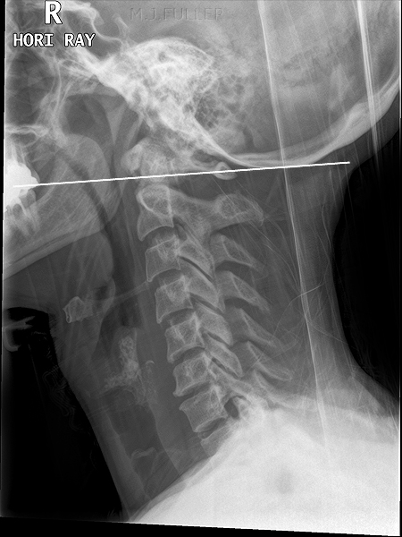
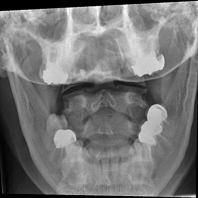
On some patients you will not be able to achieve a satisfactory demonstration of the odontoid process using the conventional AP open-mouth technique. Fuch's technique provides an alternative technique to use in patients who do not have an acute cervical spine injury.
The Occipitomental Projection
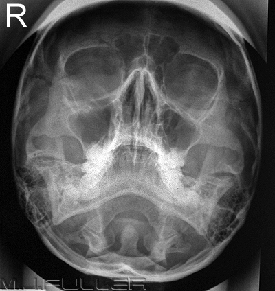
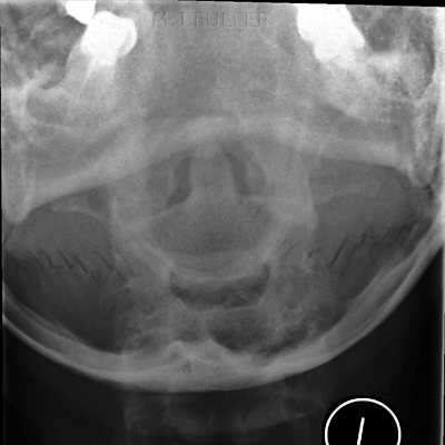
You may have noticed that you can visualise the odontoid peg on the occipito-mental (OM) image of the facial bones. A coned OM view can be considered as an alternative technique for imaging the odontoid peg. The disadvantage of this projection is that the odontoid peg and related anatomy are projected over the occiput of the skull. If you're lucky, the foramen magnum will be conveniently positioned.
Important note: extending a patient's neck to achieve an OM position is conta-indicated in a trauma patient
Case Study1
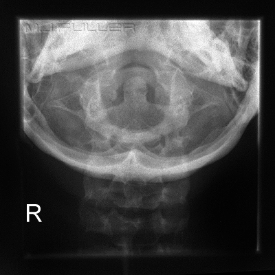
This image is a coned OM for odontoid peg (Fuch's View). The original cone marks are displayed.
A frequent failing of this view is underexposure (this image is slightly underexposed). The exposure technique should be as for an OM projection of the facial bones rather than an AP cervical spine projection.
Further information here <a class="external" href="http://bloggingradiography.blogspot.com/2007/05/odontoid-trouble.html" rel="nofollow" target="_blank">http://bloggingradiography.blogspot.com/2007/05/odontoid-trouble.html</a>
Comment
Fuch's method should not be attempted in patients with acute cervical spine injury- when in doubt..don't
Links
Further information can be found on Jeremy Enfinger's excellent radiography blog at <a class="external" href="http://bloggingradiography.blogspot.com/2007/05/odontoid-trouble.html" rel="nofollow" target="_blank">http://bloggingradiography.blogspot.com/2007/05/odontoid-trouble.html</a>
...back to the Applied Radiography home page
