Flexion and Extension Cervical Spine Radiography
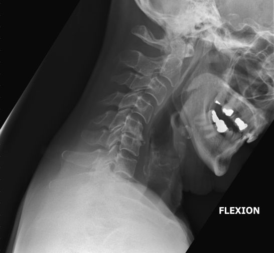
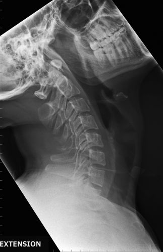
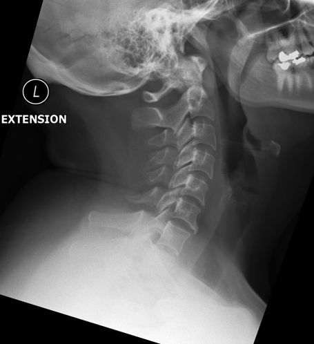
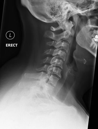
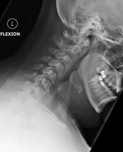
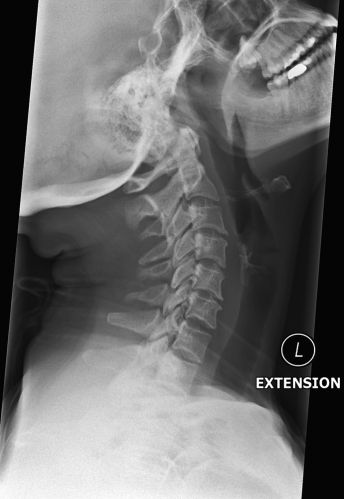
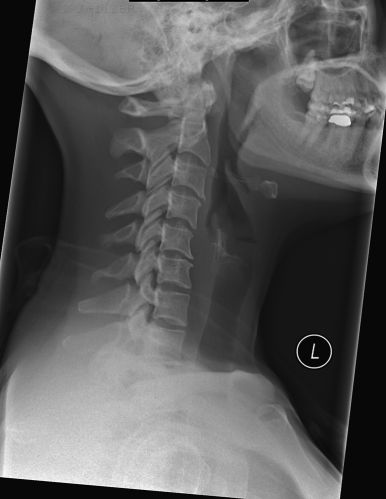
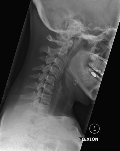
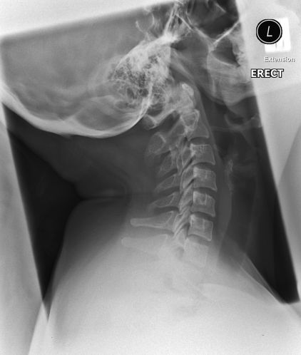
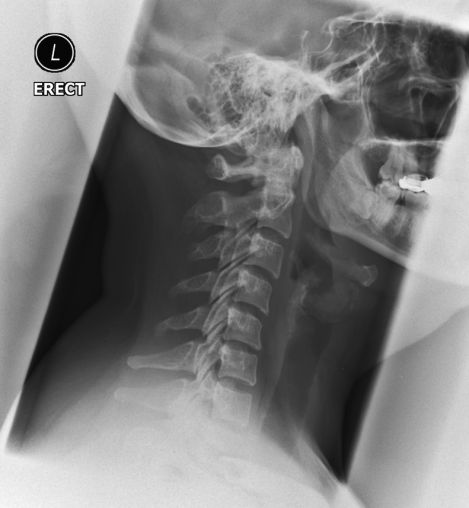
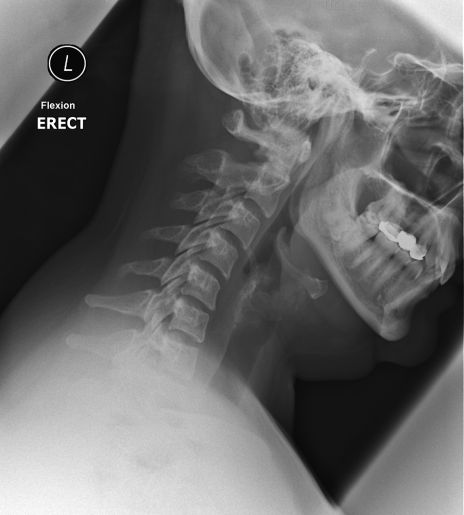
Flexion and extension functional radiography has not been universally accepted as a diagnostic tool. The advent of MRI imaging has further reduced the acceptability of radiographic dynamic imaging of the cervical spine. Despite this variability in acceptance, flexion and extension lateral cervical spine radiography is still practiced in some centres in the acute and/or follow up settings. This page considers all aspects of function cervical spine radiography.
Technique
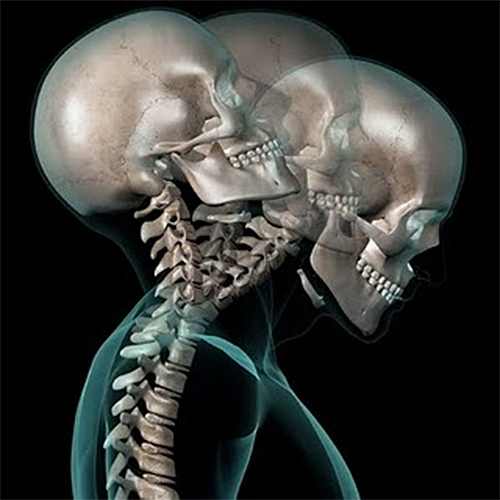
Self-limiting Cervical Movements
Flexion and extension views of the cervical spine should not be performed on patients with unstable cervical spine fractures. Under no circumstances should routine flexion and extension views be performed in an acute setting without the supervision/consent of a neuosurgeon (or other suitably qualified medical specialist). Given the risks of injury to the patient associated with functional cervical spine views, the requested functional views should be in writing (preferable from a neurosurgeon). It is good radiographic practice to review the old imaging yourself (don't rely on others) to ensure that you are not putting the patient at risk- don't rely on a verbal assurance that the cervical spine CT was normal.
An additional problem with functional views of the cervical spine in the trauma setting is that the patient may be unable or unwilling to flex and extend his/her neck. Wang et al reported the following
Flexion and extension lateral radiographs of the cervical spine may suggest signs of ligamentous and soft tissue injuries in a potentially unstable spine. However, patients with acute injuries and severe pain and muscle spasms may not be able to move their necks effectively, severely compromising the diagnostic yield of the radiographs. In addition, there are reports of serious neurologic injuries occurring with the use of these radiographs in acutely injured patients.
....Of the 290 flexion and extension radiographs, 97 (33.5%) of them showed such little or inadequate flexion or extension movement that cervical stability could not be assessed. Flexion and extension cervical radiographs should not be obtained routinely in the emergency department because 13 of these studies will be inadequate because of pain and muscle spasms experienced by patients. Patients with cervical injuries may not be able to fully flex and extend their necks; this may lead to false reassurance to patients who actually have had an inadequate study to diagnose potential instability.
Clin Orthop Relat Res. 1999 Aug;(365):111-6.
Cervical flexion and extension radiographs in acutely injured patients.
</a>
Genuine Cervical Flexion and Extension Movements
<a class="external" href="http://www.squidoo.com/whiplash-and-personal-injury-analysis" rel="nofollow" target="_blank">
http://www.squidoo.com/whiplash-and-personal-injury-analysis</a>Flexion and extension cervical spine radiography can be self defeating in the acute setting. Patients will be referred for cervical spine flexion and extension radiography because of trauma to the cervical spine. The pain from this trauma prevents the patients from flexing and extending their cervical spine. The patient may appear to be flexing and extending their cervical spine, but what they are actually doing is flexing and extending using their hips and lumbar spine.
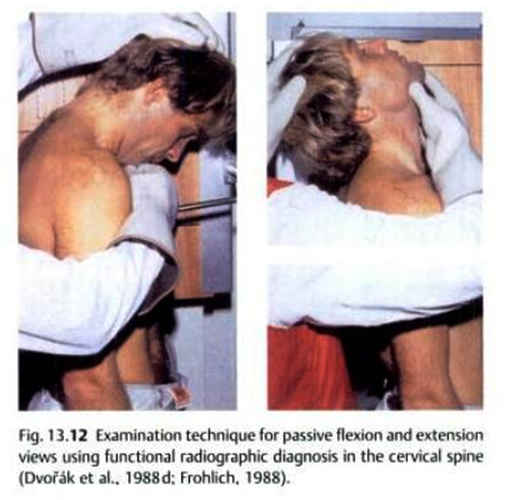
Passive Cervical Movements
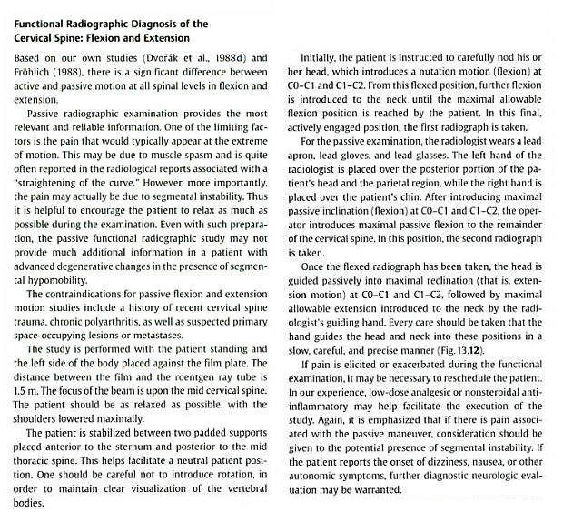
Functional radiography of the cervical spine can also be achived with passive movement of the cervical spine. Passive movement is where the flexion and extension movements are controlled by a Radiologist (or other suitably qualified health profesional) and not by the patient- the patient is passive. [Flexion and Extension Radiography&hl=en&ei=xFtTTYnAHMnCcfe1mKYH&sa=X&oi=book_result&ct=result&resnum=10&ved=0CF8Q6AEwCTge#v=onepage&q&f=false|Title Musculoskeletal manual medicine: diagnosis and treatment Author Jiří Dvořák Edition illustrated Page: 13 Publisher Thieme, 2008] I cannot overemphasize the risks of this procedure in any patient and, in particular, in a patient with a recent injury. When in doubt, seek more information before proceeding.
Case 1
Case 2
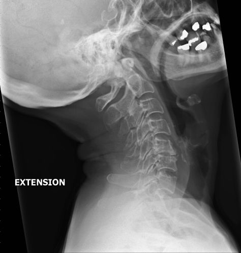
30 year old female with upper thoracic spine pain worse on cervical movement
Case 3
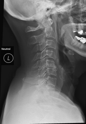
42 year old female with neckpain post RTA/MVA. There is an anterior ostoephyte at C6. There is some loss of normal cervical lordosis
Case 4
Case 5
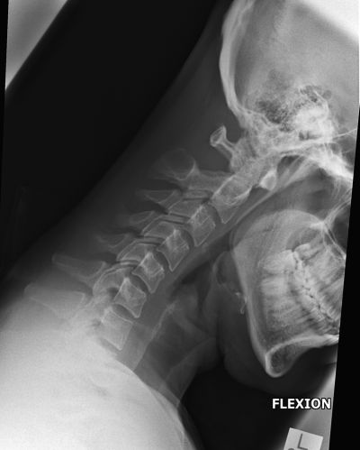
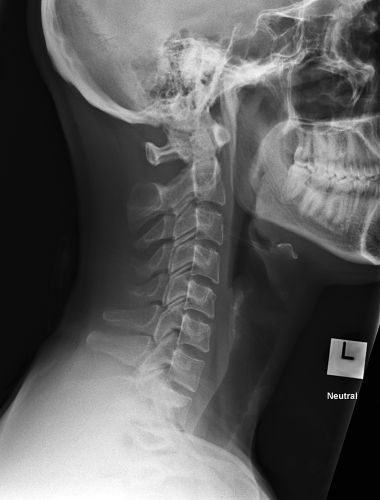
32 year old female with unknown history. There is loss of the normal cervical lordosis
... back to the wikiradiography home page
... back to the Applied Radiography home page
