Fasciae and Ligaments of the Anterior Abdominal Wall

From wikiRadiography
Revision as of 16:46, 11 November 2020 by Travis (talk | contribs) (Created page with "<div class="WPC-editableContent"><table width="100%"><tr><td><div class="WPC-edit-area"><br/><h3>Fasciae and Ligaments of the Anterior Abdominal Wall</h3><ul><li>Are organize...")
(diff) ← Older revision | Latest revision (diff) | Newer revision → (diff)
Jump to navigation Jump to search

Fasciae and Ligaments of the Anterior Abdominal Wall

  • Are organized into superficial (tela subcutanea) and deep layers; the superficial layer has a thin fatty (Camper's fascia ), and the deep layer has a membranous layer (Scarpa's fascia ).

Superficial fascia


Superficial layer of the superficial fascia (Camper's fascia)

  • Continues over the inguinal ligament to merge with the superficial fascia of the thigh.
  • Continues over the pubis and perineum as the superficial layer of the superficial perineal fascia.

Deep layer of the superficial fascia (Scarpa's fascia)

  • Is attached to the fascia lata just below the inguinal ligament.
  • Continues over the pubis and perineum as the membranous layer (Colles' fascia ) of the superficial perin fascia.
  • Continues over the penis as the superficial fascia of the penis and over the scrotum as the tunica darto which contains smooth muscle.
  • May contain extravasated urine between this fascia and the deep fascia of the abdomen, resulting from rupture of the spongy urethra.

Deep fascia

  • Covers the muscles and continues over the spermatic cord at the superficial inguinal ring as the external spermatic fascia.

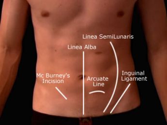
Linea alba
  • Is a tendinous median raphe between the two rectus abdominis muscles, extending from the xiphoid process to the pubic symphysis.
  • Is formed by the fusion of the aponeuroses of the external oblique, internal oblique, and transverse muscle to the abdomen.

Linea semilunaris
  • Is a curved line along the lateral border of the rectus abdominis.
Linea semicircularis (arcuate line)
  • Is a crescent-shaped line marking the inferior limit of the posterior layer of the rectus sheath just below level of the iliac crest.
Lacunar ligament (Gimbernat's ligament)
  • Represents the medial triangular expansion of the inguinal ligament to the pectineal line of the pubis.
  • Forms the medial border of the femoral ring and the floor of the inguinal canal.
Pectineal (Cooper's) ligament
  • Is a strong fibrous band that extends laterally from the lacunar ligament along the pectineal line

Inguinal ligament (Poupart's ligament)
  • Is the folded lower border of the aponeurosis of the external oblique muscle, extending between the anterior superior iliac spine and the pubic tubercle.
  • Forms the floor (inferior wall) of the inguinal canal.
Iliopectineal arcus or ligament
  • Is a fascial partition that separates the muscular (lateral) and vascular (medial) lacunae deep to the inguinal ligament.
    • The muscular lacuna transmits the iliopsoas muscle.
    • The vascular lacuna transmits the femoral sheath and its contents, including the femoral vessels, a femoral branch of the genitofemoral nerve, and the femoral canal.
Reflected inguinal ligament
  • Is formed by certain fibers of the inguinal ligament reflected from the pubic tubercle upward toward the linea alba.
  • Also has some reflection from the lacunar ligament.
Falx inguinalis (conjoint tendon)
  • Is formed by the aponeuroses of the internal oblique and transverse muscles of the abdomen and is into the pubic tubercle.
  • Strengthens the posterior wall of the medial half of the inguinal canal.


Fascia of the Abdomen

Rectus sheath

  • Is formed by fusion of the aponeuroses of the external oblique, internal oblique, and transverse muscle of the abdomen.
  • Encloses the rectus abdominis and sometimes the pyramidal muscle.
  • Also contains the superior and inferior epigastric vessels and the ventral primary rami of thoracic nerves


Anterior layer of the rectus sheath
  • Above the arcuate line: aponeuroses of the external and internal oblique muscles.
  • Below the arcuate line: aponeuroses of the external oblique, internal oblique, and transverse muscles
Posterior layer of the rectus sheath
  • Above the arcuate line: aponeuroses of the internal oblique and transverse muscles.
  • Below the arcuate line: rectus abdominis is in contact with the transversalis fascia.

◄.....Go back to the Gross Anatomy homepage