Elbow Trauma 1
This seventeen year old girl presented to the Emergency Department with a painful elbow after falling off a stool.
Imaging
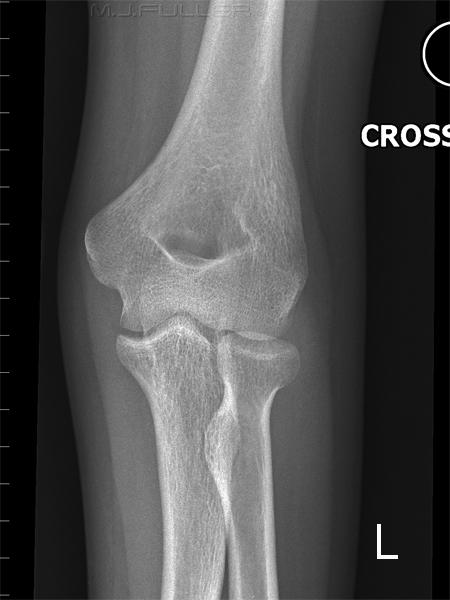
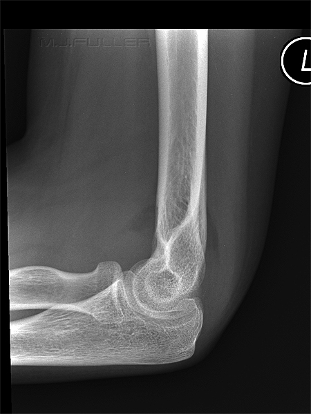
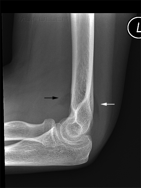
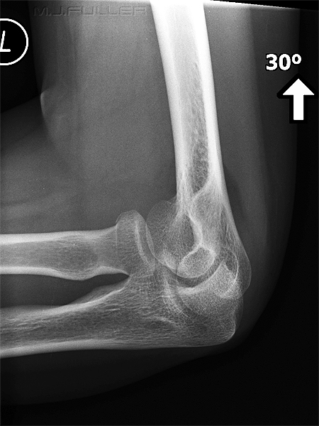
The radiographer has performed AP and lateral images of the patient's elbow as shown below.
Are there any abnormal findings? Would further views be useful?
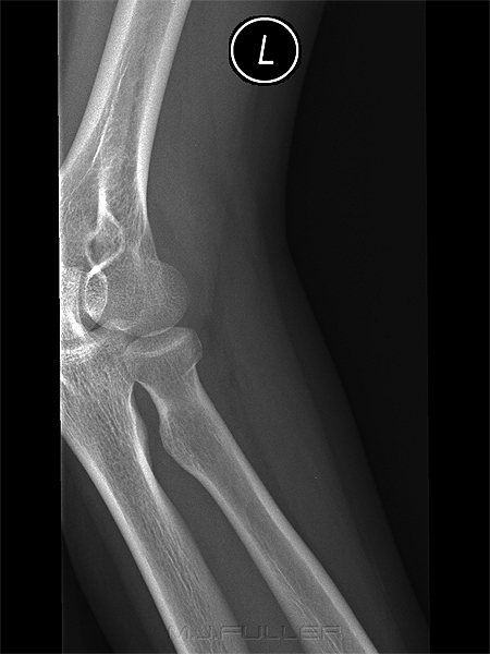
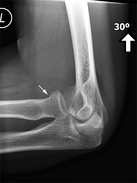
The radiographer performed an oblique elbow view in the hope of revealing a radial head fracture as shown below.
Has this projection revealed a fracture?
If not, what would you do now?
The radiographer decided that there was a very high chance that there was a radial head fracture and that it might be demonstrated with further imaging. A further elongated view of the radial head was performed
The loss of smooth contour of the radial head on this view is indicative of a radial head fracture....game over
Discussion
The radiographer has taken a logical path in the imaging that has been undertaken. You could question the value of the supplementary views when the patient was likely to be treated for a radial head fracture on clinical grounds alone. For what it's worth, I believe the pursuit of the radial head fracture was warranted. Without the additional views, you are reporting back to the referring doctor that the patient might have a radial head fracture. This is not new information... the patient was referred for radiography because it was considered that she had sustained a radial head fracture in the fall. The firm diagnosis allows the referring doctor to treat the patient with confidence. He/she is able to explain to the patient with confidence the nature of the fracture and how it should be treated. Importantly, the doctor is able to explain to the patient how long it will take to heal and advise on appropriate limitations of activity and appropriate pain relief.
....back to the Applied Radiography home page
