Dislocations and Subluxations of the Carpus
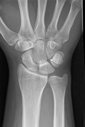
Scapholunate Instability (the Terry Thomas Sign)Subluxations and dislocations of the carpal bones are more commonly missed than fractures. I suspect that a lack of pattern recognition is a significant factor in the failure to diagnose these injuries. This page examines the more common carpal bone dislocations and subluxations
Lunate and Perilunate Dislocations
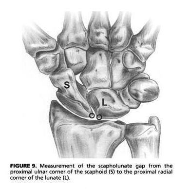
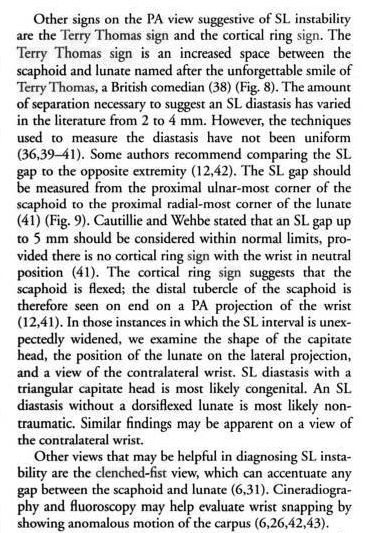
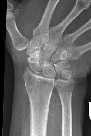
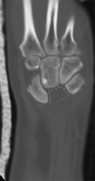
"The most common widened carpal joint space is that of the scapholunate. Nicknamed the "Terry-Thomas sign" by Frankel ...., widening at this joint has been variously defined as anything from 2 mm [Ref Moneim, JBJS 1981; Linscheid, JBJS, 1972 ] to 4 mm... Absolute magnitudes are difficult to define and more difficult to justify: the magnification of the film is rarely available. Relative widening at this joint is simple and easy to determine. As mentioned above, however, the scapholunate joint has a varying profile, from its proximal to its distal margin, due to the rounded contour of the scaphoid’s lunate facet. Additionally there is variance in the width of the scapholunate joint in its dorsal, middle and palmar aspects. ..., however, the narrowest aspect of the joint in the same coronal plane should be measured, not the most proximal part (which would make most joint spaces seem wide) or the most distal part. Cautilli and Webbe [JHS 1991] have suggested that the measurement be taken at the proximal border of these bones, and found a mean of 3.7 mm with a range from 2.5 to 5.0 mm. However, there is no control for the magnification of the film, the size of the patient, and they note that "the scaphoid’s proximal pole is rounded, making measurement more difficult." Given the inaccuracies inherent in their method, and the built-in control and ease of definition of the standard method, we recommend the scapholunate gap be measured as the narrowest gap between the two bones when measured in the midpoint of adjacent parallel articular contours. Optimally this measurement should be made on a view profiling the scapholunate joint, often with fluoroscopic control ....
Dr. Frankel graciously obtained the actor’s permission before naming a pathological entity after him. Readers not familiar with the comic actor’s famous diastasis in his front teeth are referred to Dr. Frankel’s article. Mr. Terry-Thomas kindly provided two pictures of himself for publication in CORR. The joint space must be properly profiled in order to make a precise determination of its width. This may be more difficult that it first appears, because often the joint line of interest is not properly profiled. Joint lines that are apparently well-profiled may not be so, upon closer examination.
...Fluoroscopic evaluation of the wrist between radial and ulnar deviation in both supine and prone positions allows optimal evaluation of the scapholunate joint space. The suspected widened scapholunate joint space must be compared to the opposite side to exclude the occasional borderline normally wide scapholunate joint space."<a class="external" href="http://www.hand-therapy.com/images/Radiology_100500.pdf" rel="nofollow" target="_blank">RADIOLOGY OF THE WRIST
Presented by Karen Nugent, PT, CHT and David Nelson, M.D.
At the 23rd Annual Meeting of the American Society of Hand Therapists
5 October 2000</a><a class="external" href="http://www.hand-therapy.com/images/Radiology_100500.pdf" rel="nofollow" target="_blank">
</a>
Measuring the Scapholunate Distance
<a class="external" href="http://books.google.com.au/books?id=b7-kkx-8eqYC&pg=PA485&lpg=PA485&dq=terry+thomas+sign+clenched+fist&source=bl&ots=DqTjyisJXc&sig=_yThM9e-Svrf0kHRks30TxkCnRA&hl=en&ei=DOLdSffGBtSJkQXn3LmrDA&sa=X&oi=book_result&ct=result&resnum=6#PPA485,M1" rel="nofollow" target="_blank">By Richard A. Berger, Arnold-Peter C. Weiss. Hand surgery</a>
quoted from<a class="external" href="http://books.google.com.au/books?id=b7-kkx-8eqYC" rel="nofollow" target="_blank">Hand surgery</a><a class="external" href="http://books.google.com.au/books?id=b7-kkx-8eqYC" rel="nofollow" target="_blank">By Richard A. Berger, Arnold-Peter C. Weiss</a><a class="external" href="http://books.google.com.au/books?id=b7-kkx-8eqYC" rel="nofollow" target="_blank">Edition: illustrated</a><a class="external" href="http://books.google.com.au/books?id=b7-kkx-8eqYC" rel="nofollow" target="_blank">Published by Lippincott Williams & Wilkins, 2003</a>
Radiography
The PA Clenched Fist View - used to evaluate widening of scapholunate interval;
- clenching first draws the capitate proximally & emphasizes any widening of the scapho-lunate interval;
- should get companion views of the opposite side for comparison;
- best taken: with supinated clenched fist view with wrist in ulnar deviation;
- AP X-rays made with wrist in ulnar deviation (increases gap), in radial deviation (decreases gap), & with application of longitudinal compressive load (clenched fist) may also show widened scapholunate gap in wrist with SLD;
- Technique:
- same technique as <a class="external" href="http://www.wheelessonline.com/ortho/posterior_anterior_view_of_the_wrist" rel="nofollow" target="_blank">PA view</a> of wrist except patient clenches fist as tightly as possible during exposure;
- proper technique: for visualizing scapholunate interval;
- gap is more noticeable on AP View that is made with wrist supinated than it is on more usual PA view (made with wrist pronated);
adapted from
<a class="external" href="http://www.wheelessonline.com/ortho/clenched_fist_ap" rel="nofollow" target="_blank">Wheeless' Textbook of Orthopaedics</a>
Plain Film Appearance
- Findings in Scapholunate Dissociation
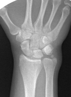
- scapholunate interval > 2-3 mm (Terry Thomas sign);
- cortical ring sign:
- produced by cortex of distal pole of palmar flexed scaphoid;
- produced by cortical outline of the distal pole of scaphoid;
- scaphoid is foreshortened;
- distance between scaphoid ring & proximal pole is < 7 mm;
- negative ulnar variance:
- misc:
- ulnar deviation, increases scapholunate gap;
- radial deviation, closes gap;
quoted from<a class="external" href="http://www.wheelessonline.com/ortho/clenched_fist_ap" rel="nofollow" target="_blank">Wheeless' Textbook of Orthopaedics</a>
Anatomy
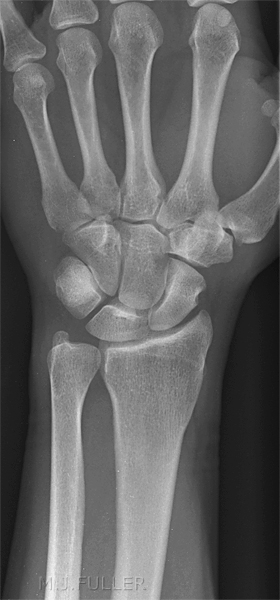
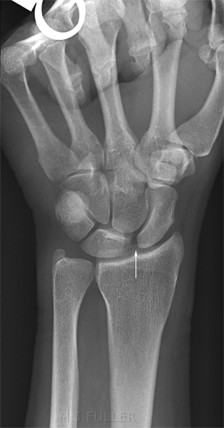
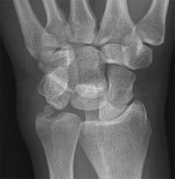
This is a type I lunate. Note that the lunate does not articulate directly with the hamate. This is a type II lunate- the hamate articulates directly with the lunate.
Case Studies
---- Case 1 ----
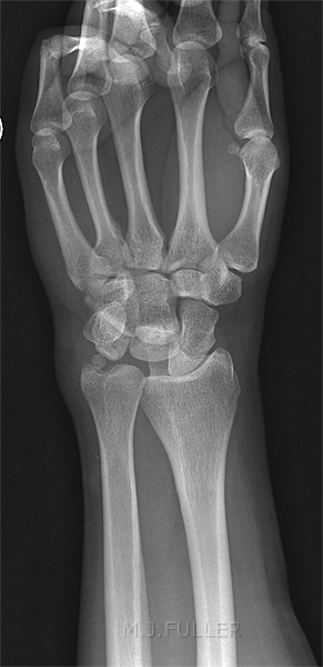
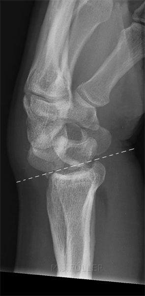
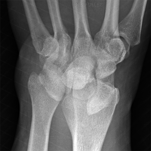
This 32 year old man presented to the Emergency Department after falling from a ladder. He was referred for an extensive list of radiographic examinations including left wrist. This is the first image from this particular trauma series. (in cases of serious trauma, the wrist imaging would not be a high priority)
There is loss of parallelism of the carpal bones. The ulnar styloid suggests a normal variant or old injury. There is loss of the normal proximal and distal carpal arc lines
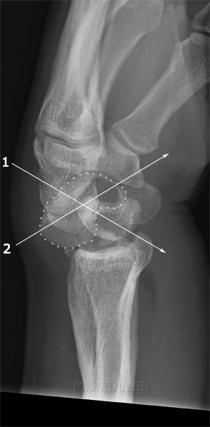
The loss of normal carpal arcs was noted by the radiographer who proceeded to perform a lateral wrist view.
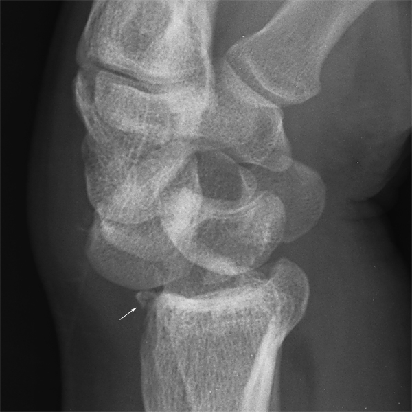
The radiographer paid particular attention to the integrity of the scaphoid.
The patient proceeded to CT followed by the operating theatre for reduction of the carpal bone dislocation.
<embed allowfullscreen="true" height="350" src="http://widget.wetpaintserv.us/wiki/wikiradiography/widget/youtubevideo/f993d65f25fd25ce84c3c26323dcfad82cfa9d41" type="application/x-shockwave-flash" width="425" wmode="transparent"/>
(Note: Youtube videos are displayed at lower resolution when 'hot-linked'. Click on the bottom right corner of the video and it will open at full resolution.)<embed allowfullscreen="true" height="350" src="http://widget.wetpaintserv.us/wiki/wikiradiography/widget/youtubevideo/02742b95ce1461aa92b5006e876262374bf8615f" type="application/x-shockwave-flash" width="425" wmode="transparent"/>
(Note: Youtube videos are displayed at lower resolution when 'hot-linked'. Click on the bottom right corner of the video and it will open at full resolution.)<embed allowfullscreen="true" height="350" src="http://widget.wetpaintserv.us/wiki/wikiradiography/widget/youtubevideo/090a5bf2d7ce4a317d6744d1f7123ae88639ed78" type="application/x-shockwave-flash" width="425" wmode="transparent"/>
(Note: Youtube videos are displayed at lower resolution when 'hot-linked'. Click on the bottom right corner of the video and it will open at full resolution.)
This is the post-reduction image from the mobile image intensifier. Comment
This case demonstrates a thoughtful approach to the radiographic demonstration of this patient's carpal injury. The radiographer recognised the carpal dislocation from the PA image. The radiographer was mindful of the importance of obtaining a good lateral projection. Equally, the radiographer paid careful attention to the imaging of the scaphoid (given the association between this carpal dislocation and scaphoid fracture). Importantly, the surgical approach in cases where a scaphoid fracture is demonstrated is different to a simple closed reduction of a perilunate dislocation.
The radiographer also noted the negative palmar tilt and considered the possibility of an old wrist fracture. This was confirmed by the patient and this information was relayed in person by the radiographer to the reporting radiologist.
This case supports the contention that trauma radiography is a specialised area of plain film radiography. In addition to all of the above, the radiographer relayed the findings in person to the referring doctor in the ED. This facilitated a timely undertaking of orthopaedic review and subsequently CT imaging of the wrist and surgical reduction. This type of personal communication helps to engender a co-operative relationship, sense of common purpose, and camaraderie between the radiographer and the referring doctor.
Finally, I would suggest that the satisfaction experienced by the radiographer in successfully completing this examination would be vastly different to a radiographer who simply undertook AP and lateral views of the patient's wrist and was unaware that the patient had sustained a significant wrist injury.
... back to the Home page
... back to the Applied Radiography home page
