Common Abdominal Pathologies and Normal Anatomical Variants

From wikiRadiography
Revision as of 16:41, 11 November 2020 by Travis (talk | contribs) (Created page with "<div class="WPC-editableContent"><font size="5"><b>Introduction</b></font><br/><blockquote>It is important in all radiography to be able to distinguish between normal anatomic...")
(diff) ← Older revision | Latest revision (diff) | Newer revision → (diff)
Jump to navigation Jump to search
Introduction

It is important in all radiography to be able to distinguish between normal anatomical variants and pathologies. This page is dedicated to comonly seen pathologies and normal anatomial variants demonstrated on abdominal plain films.


Case 1
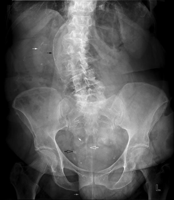
supine abdomenThere are multiple metallic surgical slips (solid white arrows)

There is a calcified segment of abdominal aorta ? aneurysmal (solid black arrow)

There are multiple pelvic phleboliths (hollow black arrow)

There is sacral spinibifida occulta (white hollow arrow)

There are abdominal hernia(s) (grey arrow)