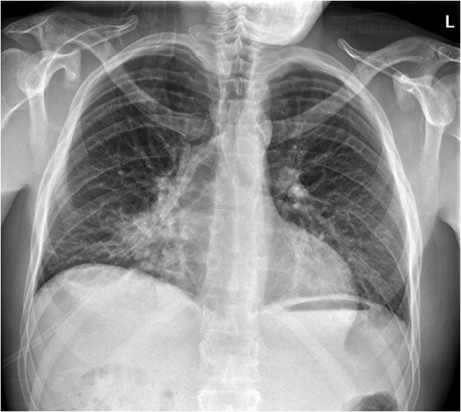
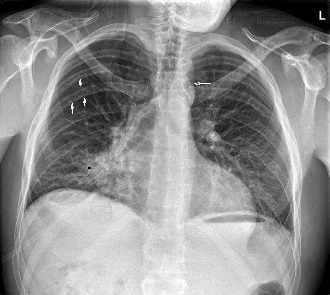
Coarctation of the Aorta
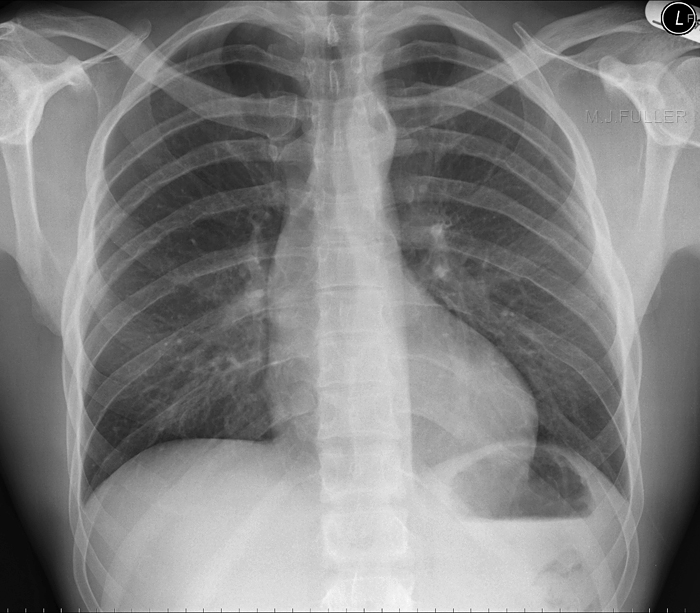
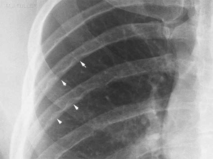
Rib NotchingCoarctation of the aorta is often identified as an incidental finding following chest radiography. Radiographers who are familiar with the plain film appearances of coarctation of the aorta have the potential to raise this frequently missed finding with the referring practitioner/radiologist.
Rib notching on palin film chest is considered pathognomonic of coarctation of the aorta. Coarctation is the most common cause of rib notching but not the only cause.
Pathology
<a class="external" href="http://en.wikipedia.org/wiki/Coarctation_of_the_aorta" rel="nofollow" target="_blank">http://en.wikipedia.org/wiki/Coarctation_of_the_aorta</a>
"Coarctation of the aorta, or aortic coarctation, is a congenital condition whereby the aorta narrows in the area where the ductus arteriosus (ligamentum arteriosum after regression) inserts"
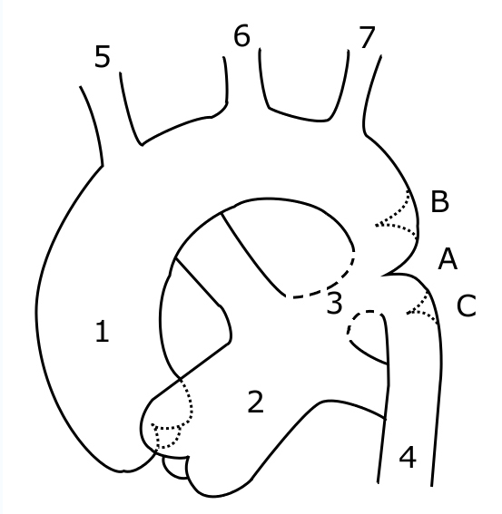
Schematic drawing of alternative locations of a coarctation of the aorta, relative to the ductus arteriosus.
A: Ductal coarctation,
B: Preductal coarctation,
C: Postductal coarctation.
1: Ascending Aorta,
2: Pulmonary Artery,
3: Ductus arteriosus,
4: Descending Aorta,
5: Brachiocephalic Artery,
6: Common Carotid Artery,
7: Subclavian Artrey<a class="external" href="http://en.wikipedia.org/wiki/Coarctation_of_the_aorta" rel="nofollow" target="_blank">http://en.wikipedia.org/wiki/Coarctation_of_the_aorta</a>
Plain Film Appearances
Case 2
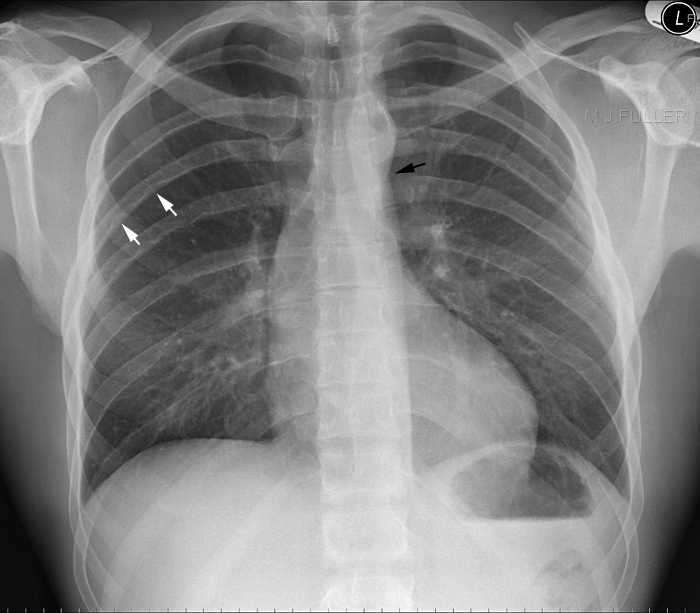
The Figure 3 Sign
It has been suggested that the figure 3 sign could be caused by
- a dilated Left Subclavian Artery
- a dilated aortic knob,
- or the “tuck” of coarct itself, and poststenotic dilatation
<a class="external" href="http://www.learningradiology.com/archives04/COW+128-Coarctation/coarctcorrect.htm" rel="nofollow" target="_blank">www.learningradiology.com</a>
These are referred to as a single sign but are infact 3 different entities.
