Chest Radiography for Inhaled Foreign Body
Introduction
Radiographic TechniquesIf you are employed in an acute care facility you will occasionally see patients with a history of suspected foreign body inhalation. If your institution caters for paediatric patients, this will be a familiar history. The radiographic techniques are specific and occasionally very effective. (Tip- don't feed young children peanuts)
Inspiration and Expiration Technique
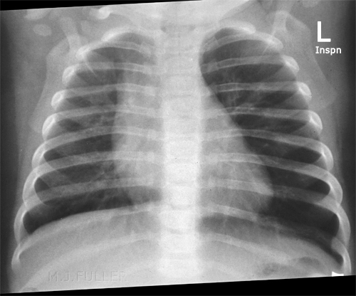
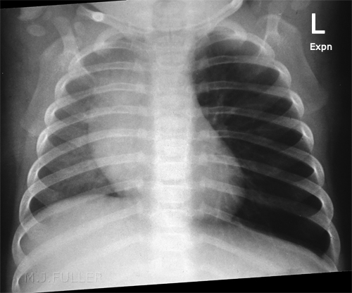
Inspiration and expiration PA/AP chest radiography is a commonly employed series for inhaled foreign body radiography in children. If an inhaled foreign body is present it can produce a one-way or ball-valve effect. This will tend to over-expand the lung distal to the foreign body. The over-expanded lung will tend to remain a constant volume on inspiration, expiration and decubitus views/techniques.
Dr Helen Williams noted the following in respect of inhaled foreign bodies. "An inhaled foreign body (FB) in a child can be easily missed if the diagnosis is not considered, particularly if a history of choking or coughing is not forthcoming, or the episode of aspiration is unwitnessed. Once the diagnosis is suspected a chest radiograph (CXR) is invariably requested. This may provide clues to the diagnosis but a normal CXR does not rule out FB aspiration. Radio-opaque foreign bodies are easily localised, usually within a major airway. However, it can be difficult to identify the radiographic signs associated with an inhaled FB that is not radio-opaque—for example, food or small plastic objects. The presence of an intraluminal FB within the trachea or a bronchus often results in secondary changes in the associated lung or pulmonary lobe. One of the most important signs to identify is obstructive emphysema, or overinflation of the lung or lobe distal to the airway obstruction"
<a class="external" href="http://ep.bmjjournals.com/cgi/content/extract/90/2/ep31" rel="nofollow" target="_blank">Dr Helen Williams
ILLUMINATIONS </a><a class="external" href="http://ep.bmjjournals.com/cgi/content/extract/90/2/ep31" rel="nofollow" target="_blank"> Inhaled foreign bodies </a>
<a class="external" href="http://ep.bmjjournals.com/cgi/content/extract/90/2/ep31" rel="nofollow" target="_blank">http://ep.bmjjournals.com/cgi/content/extract/90/2/ep31</a>
Decubitus Technique
Jayesh M Bhatt, VikkiAnn Lee, Penny Broadley commented on Dr Helen Williams article as follows "Williams [1] highlights the key points in the management of inhaled foreign bodies (FB). One of the important points is to request inspiratory and expiratory chest radiographs if FB aspiration is suspected. Because young children are unable to co-operate, paired inspiratory and expiratory films are not always possible. The lateral decubitus film is a useful and convenient method to determine the presence of air trapping [2] a... When the child is placed on his side, the splinting of the dependent hemithorax results in restriction of movement of the thoracic cage on that side and under aeration of the lung while the hemithorax on the opposite side is unrestricted and the lung is well aerated"
<a class="external" href="http://ep.bmj.com/cgi/eletters/90/2/ep31#44" rel="nofollow" target="_blank">Jayesh M Bhatt, VikkiAnn Lee, Penny Broadley</a><a class="external" href="http://ep.bmj.com/cgi/eletters/90/2/ep31#44" rel="nofollow" target="_blank">
The lateral decubitus film to determine the presence of air trapping
http://ep.bmj.com/cgi/eletters/90/2/ep31#44
BMJ. 2 August 2005</a>
Plain Film Appearances of Inhaled Foreign Body
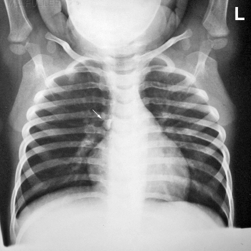
This 7 month old boy presented to the Emergency Department after a choking episode. There was concern about an inhaled foreign body. Inspiration and expiration chest radiography was requested.
Case 1
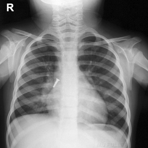
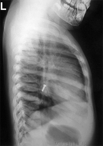
This case is circa 1958. The details of the case are unknown.
There is a bolt in the right main bronchus The bolt appears to have moved distally into a RLL bronchus.
Case 2
The details of the case are unknown.
There is a piece of gravel in the right main bronchus.(arrowed)
...back to the Wikiradiography home page
...back to the Applied Radiography home page
