Chest Normal Anatomical Variants

From wikiRadiography
Revision as of 16:39, 11 November 2020 by Travis (talk | contribs) (Created page with "<div class="WPC-editableContent"><font size="5"><b>Introduction</b></font><br/><blockquote>There are a number of normal anatomical variants visible on chest X-ray images that...")
(diff) ← Older revision | Latest revision (diff) | Newer revision → (diff)
Jump to navigation Jump to search
Introduction

There are a number of normal anatomical variants visible on chest X-ray images that can mimic pathology. This page considers some of the more commonly occuring normal chest anatomical variants that might cause confusing appearances on chest X-ray images.

Cervical Rib
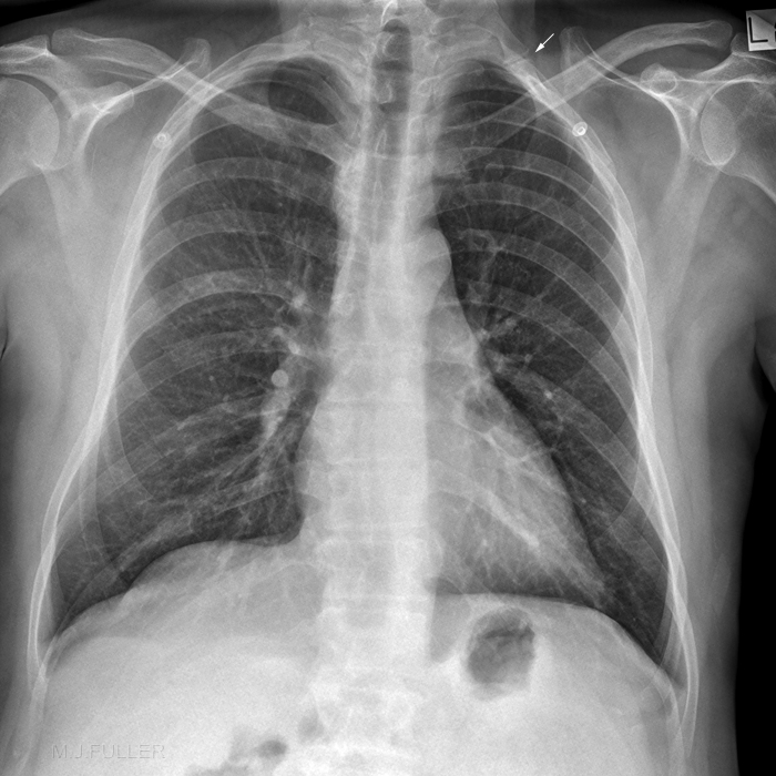
cervical ribA cervical rib is a supernumerary rib which arises from a cervical vertebra (usually the seventh).

This patient has a left sided cervical rib (arrowed). The cervical rib is largely superimposed over the second rib.


Pectus Excavatum

Case 1

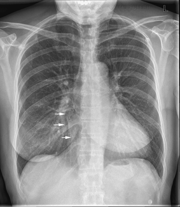
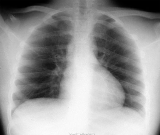
pectus excavatumThis patient presented for chest radiography with a history of recent facial nerve palsy. There is loss of the right cardiac border.
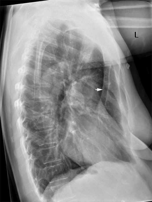
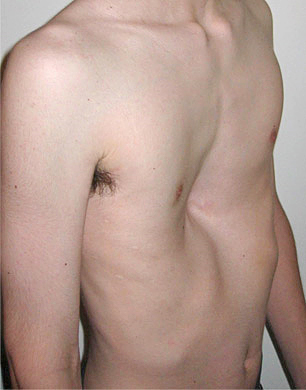
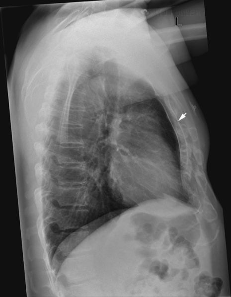
pectus excavatumThe lateral chest X-ray image demonstrates a severe pectus excavatum deformity (arrowed). In these patients the heart tends to be displaced towards the left as a result of the limited space between the depressed sternum and the spine. The loss of the right heart border on the PA image was not a result of a disease process.

Pectus Excavatum
pectus excavatum
<a class="external" href="http://en.wikipedia.org/wiki/File:Pectus1.jpg" rel="nofollow" target="_blank">http://en.wikipedia.org/wiki/File:Pectus1.jpg</a>


Pericardial fat
pericardial fat
<a class="external" href="http://jcmr-online.com/content/11/1/15" rel="nofollow" target="_blank">Validation of cardiovascular magnetic resonance assessment of pericardial adipose tissue volume</a>
<a class="external" href="http://jcmr-online.com/content/11/1/15" rel="nofollow" target="_blank">Adam J Nelson , Matthew I Worthley , Peter J Psaltis , Angelo Carbone , Benjamin K Dundon , Rae F Duncan , Cynthia Piantadosi , Dennis H Lau , Prashanthan Sanders , Gary A Wittert and Stephen G Worthley</a><a class="external" href="http://jcmr-online.com/content/11/1/15" rel="nofollow" target="_blank">Cardiovascular Research Centre, Royal Adelaide Hospital & Disciplines of Medicine and Physiology, University of Adelaide, Adelaide, SA, Australia</a>
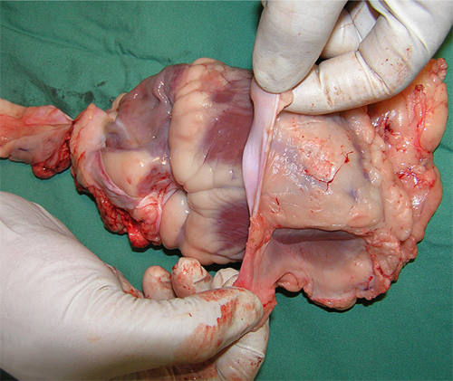
This is a sheep's heart with the pericardium partly dissected off. Note the pericardial fat.


Case 1

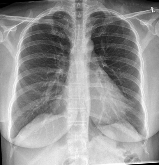
false consolidationThis 49 year old female patient presented for chest radiography with "recent onset of chest pain".

There is loss of clarity of the right heart border. In addition, there is loss of clarity of the left heart border. There are good reasons to consider that these silhouette signs are not caused by lung consolidation.
  • The patients history and clinical signs do not suggest a diagnosis of infection/consolidation
  • there is no evidence of alveolar opacity
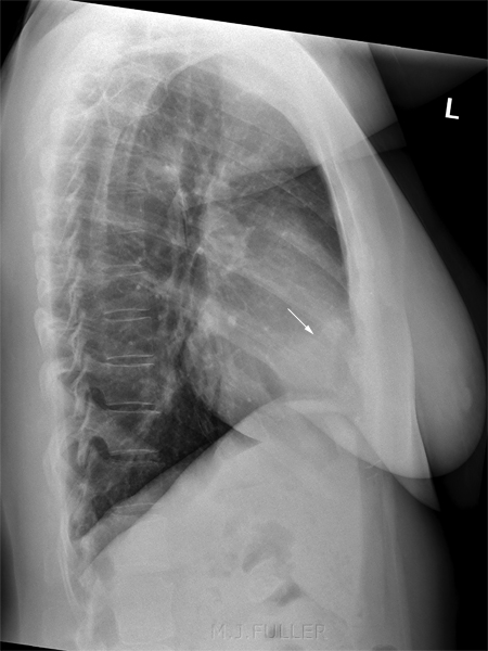
  • the lateral view does not demonstrate alveolar opacity that you would expect with a lung consolidation
The likely cause of the loss of clarity of the cardiac silhouette is the presence of pericardial fat.
false consolidation
There is no abnormal RML or left upper lobe (lingula segment) opacity. Where there is a significant amount of pericardial fat visible on the PA/AP chest image, you would expect to see evidence of this on the lateral image (white arrow)
pectus excavatum
patient with minimal pericardial fat for comparison (ignore arrow)


Dextracardia
Dextracardia in the Resus Room - wikiRadiography
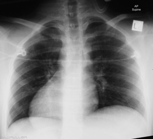
This patient has arrived at a major trauma hospital after transfer from a rural hospital. This patient has sustained unknown injuries from a motor vehicle accident (MVA). The patient has arrived with radiographs taken at the previous hospital. These radiographs include the chest image shown above. There is life-threatening danger associated with this image- what is missing?
There are several shortcomings evident in the image above. Two of the most important are that it does not have a side marker and it has no patient name or other patient details. Given the failings of this image, the chest X-ray examination was repeated in the resuscitation room as shown above. The patient has dextracardia which is a normal anatomical variant in which there is left to right reversal of the normal chest viscera.


... back to the Applied Radiography home page