Case of the week
Jump to navigation
Jump to search
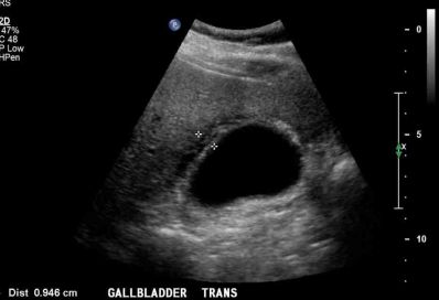
Case 1: 19 yr old male with RUQ abdominal pain + fever
Image 1 long view Rt liver 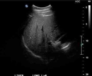 | Image 2 long gallbladder | |
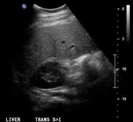
Image 3 long gallbladder 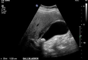 | Image 4 Liver trans | . |
Image 5 II theatre film 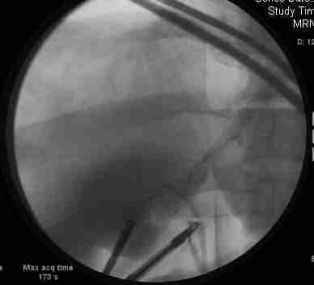 |
Answers at bottom of page
_______________________________________________________________________
Case2 - 30 yr old man eye ultrasound
Image 1 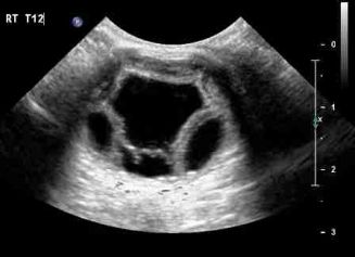 | Image 2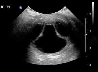 |
Answers at bottom of page
________________________________________________________________________________________________________
Case 3
Image 1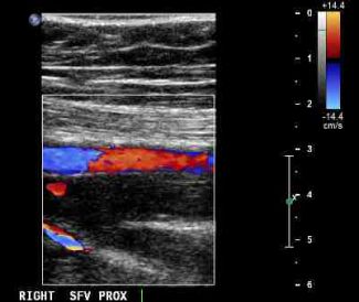 | Image 2 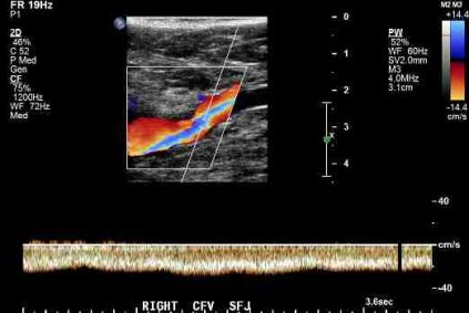 |
Image 3 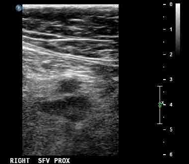 | Image 4 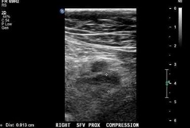 |
Answers Below
____________________________________________________________________________________________________Case1
Acute cholecystitis, stone impacted in Gb neck.thick walled gb with pericholycystic oedema.
Slight fatty infiltration of the liver.
Case 2
Eye u/s
Choroidal effusion with nodular retinal detachment
Case 3
DVT involving Femoral vein and deep femoral vein.
Go back to US main page
