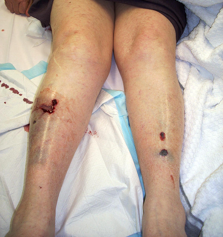
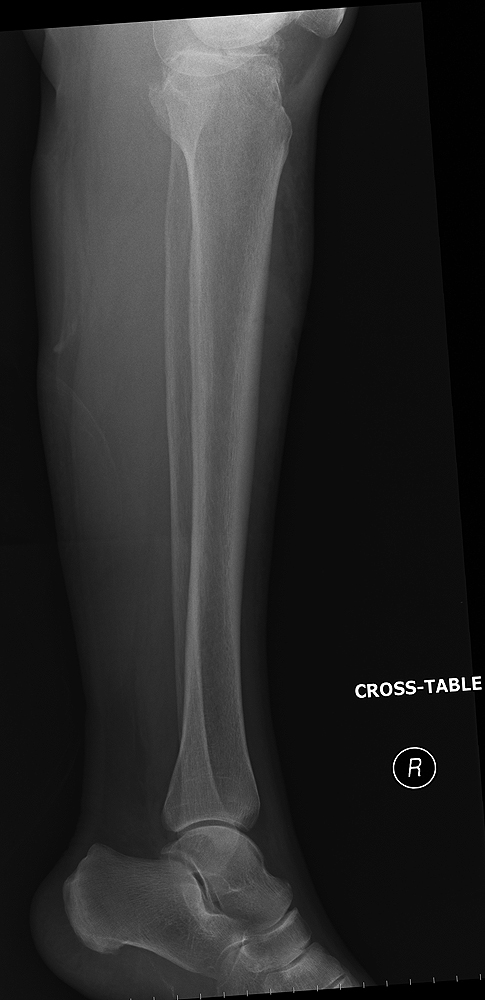
This 79 year old lady presented to the Emergency Department following a fall. She was examined and found to have a painful right lower leg with an anterior skin tear/laceration and bruising. She reported having extreme difficulty weight-bearing on her right leg following the fall. She was referred for right lower leg radiography.
Her left leg was also injured in the fall but was thought to be a soft tissue injury only.
Her lower legs and initial plainfilm imaging is shown below.
Question 1
Is there evidence of bony injury or joint dislocation demonstrated on the plain films?
Question 2
Does the clinical history/patient presentation provide any indications of possible bone/joint injury?
Question 3
What additional imaging (if any) is warranted?
wikiradiography members can provide a response using the thread at the bottom of the page
Answer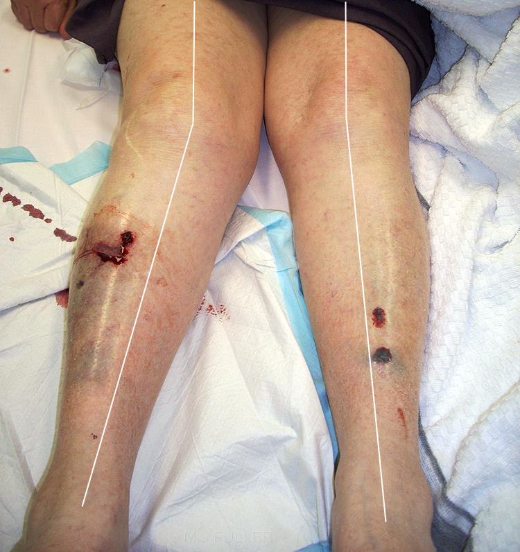 | The radiographer considered the patient's age, sex , mechanism of injury and clinical signs to indicate possible bony injury. Females over the age of 60 years are at high risk of osteoporosis predisposing them to bony injury.
The radiographer noted a difference in alignment between the patient's knees (Q-angle). Whilst this could be due to knee joint degenerative disease, a tibial plateau fracture would also account for the appearance.
Note that at this stage, the radiographer has only asked the patient her about her injury and clinical signs. Despite the fact that no imaging has been undertaken, the radiographer is already compiling a list of differential diagnoses. Whilst this might appear unimportant, a targeted search will be undertaken by the radiographer to ensure that subtle signs of tibial plateau fracture are not overlooked on the initial imaging. |
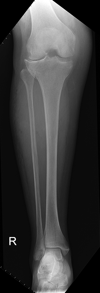 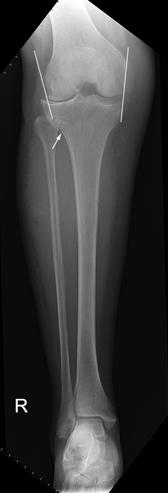 | The AP tib/fib image demonstrated two important findings
- there is a cortical irregularity of the lateral aspect of the proximal tibial metaphysis
- there is abnormal alignment of the knee joint articular surfaces laterally
At this point the radiographer considered that the patient has sustained a lateral tibial condyle fracture during her fall. The objective at this point is to confirm the existence of the fracture unequivocally and to demonstrate the nature and extent of the fracture. |
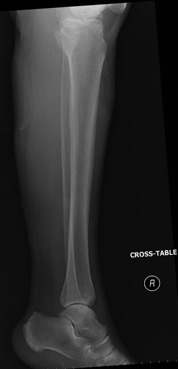 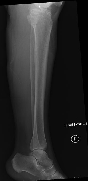 | The lateral tib/fib projection provides little by way of new information. There is a subtle cortical breach of the proximal tibial articular surface (arrowed) |
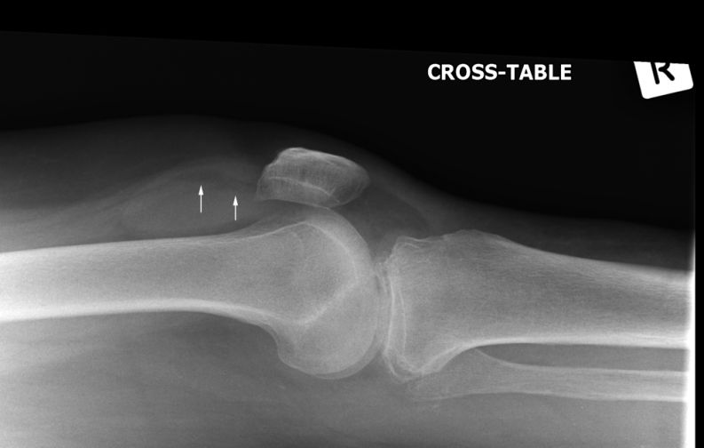 | The horizontal ray lateral knee projection image demonstrates a large knee effusion with a superior fat density indicating lipohaemarthrosis. |
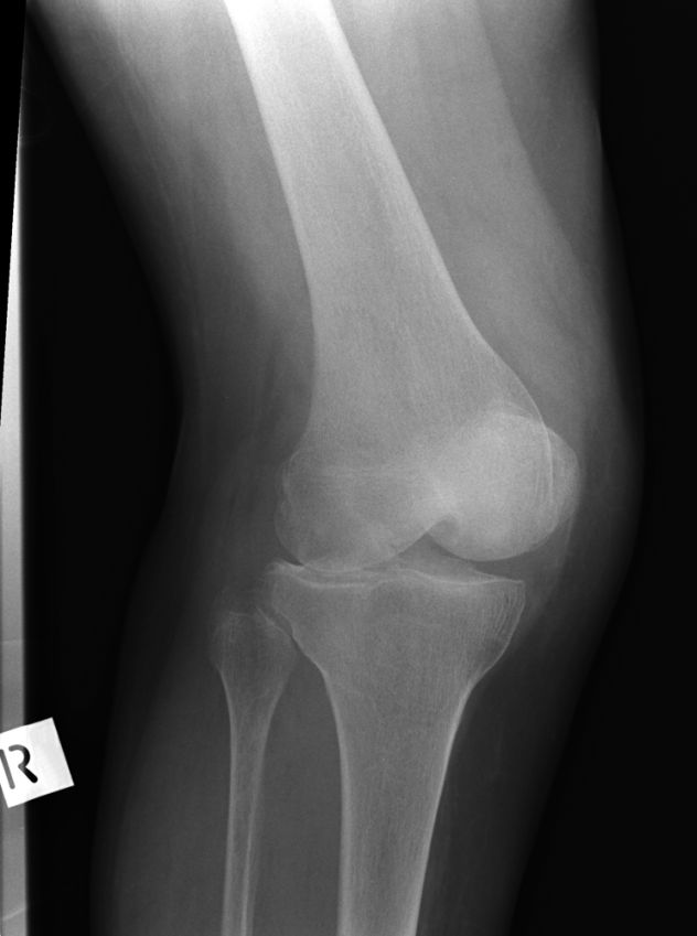 | Oblique projections of the knee can be very useful in demonstrating tibial plateau fractures. The radiographer performed both oblique projections of the patient's right knee.
Some cortical irregularity of the proximal tibial metaphysis and articular surface is demonstrated, but it is subtle and unconvincing of an acute fracture. |
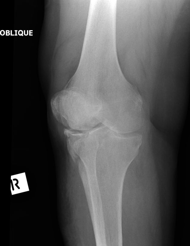
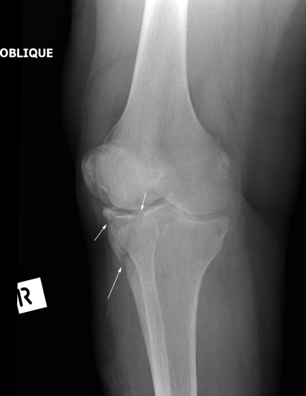 | The external oblique position demonstrates the fracture clearly. |
Discussion
The radiographer has demonstrated the patient's tibial fracture by employing a clinical approach to radiography. The radiographer's aim is not simply to produce images of the patient's lower leg, but to take on shared responsibility for establishing the correct diagnosis (MDT thinking). It could be argued that if it was known that the patient had sustained a tibial fracture, and a knee CT scan would be definitely be performed, supplementary plain film imaging was not warranted.
