Artefacts
Jump to navigation
Jump to search
An artefact is ...
Patients are normally asked to take off removable metal objects such as jewelry before scanning commences. For non-removable items, such as dental fillings, prosthetic devices, and surgical clips, it is sometimes possible to use gantry angulation to exclude the metal inserts from scans of nearby anatomy. When it is impossible to scan the required anatomy without including metal objects, increasing technique, especially kilovoltage, may help penetrate some objects, and using thin sections will reduce the contribution due to partial volume artifact.
Streaking caused by over-ranging can be greatly reduced by means of special software corrections. Manufacturers use a variety of interpolation techniques to substitute the over-range values in attenuation profiles.
The usefulness of metal artifact reduction software is sometimes limited because, although streaking distant from the metal implants is removed, there still remains a loss of detail around the metal-tissue interface, which is often the main area of diagnostic interest. Beam hardening correction software should also be used when scanning metal objects to minimize the additional artifacts due to beam hardening.
Article from: <a class="external" href="http://radiographics.rsna.org/content/24/6/1679.full" rel="nofollow" target="_blank">http://radiographics.rsna.org/content/24/6/1679.full</a><a class="external" href="http://radiographics.rsna.org/content/24/6/1679.full" rel="nofollow" target="_blank">
</a>
 e special features on some scanners designed to minimize the resulting artifacts. Avoidance of Motion Artifacts by the Operator.— The use of positioning aids is sufficient to prevent voluntary movement in most patients. However, in some cases (eg, pediatric patients), it may be necessary to immobilize the patient by means of sedation. Using as short a scan time as possible helps minimize artifacts when scanning regions prone to movement. Respiratory motion can be minimized if patients are able to hold their breath for the duration of the scan. The sensitivity of the image to motion artifacts depends on the orientation of the motion. Therefore, it is preferable if the start and end position of the tube is aligned with the primary direction of motion, for example, vertically above or below a patient undergoing a chest scan. Specifying body scan mode, as opposed to head scan mode, may automatically incorporate some motion artifact reduction in the reconstruction. Built-in Features for Minimizing Motion Artifacts.— Manufacturers minimize motion artifacts by using overscan and underscan modes, software correction, and cardiac gating. Overscan and underscan modes: The maximum discrepancy in detector readings occurs between views obtained toward the beginning and end of a 360° scan. Some scanner models use overscan mode for axial body scans, whereby an extra 10% or so is added to the standard 360° rotation. The repeated projections are averaged, which helps reduce the severity of motion artifacts. The use of partial scan mode can also reduce motion artifacts, but this may be at the expense of poorer resolution. Software correction: Most scanners, when used in body scan mode, automatically apply reduced weighting to the beginning and end views to suppress their contribution to the final image. However, this may lead to more noise in the vertical direction of the resultant image, depending on the shape of the patient. Additional, specialized motion correction is available on some scanners. Cardiac gating: The rapid motion of the heart can lead to severe artifacts in images of the heart and to artifacts that can mimic disease in associated structures, for example, dissected aorta. To overcome these difficulties, techniques have been developed to produce images by using data from just a fraction of the cardiac cycle, when there is least cardiac motion.
Article from: <a class="external" href="http://radiographics.rsna.org/content/24/6/1679.full" rel="nofollow" target="_blank">http://radiographics.rsna.org/content/24/6/1679.full</a>
The relationship between helical pitch and the severity of helical artefacts is more complex on multi-slice than on single-slice scanners. Artefacts appear to be slightly reduced when non-integer pitch values, relative to detector acquisition width, are employed. This is because z-axis sampling density is maximised for noninteger pitches.
From GE: <a class="external" href="http://www.medcyclopaedia.com/library/topics/volume_i/p/partial_volume_effect.aspx" rel="nofollow" target="_blank" title="GE Medcyclopaedia">GE Medcyclopaedia</a>
Beam Hardening Artefacts
Beam hardening artefacts appear as streaks and shadows adjacent to areas of high density such as the petrous bones, shoulders, and hips. The artefact occurs because the high density anatomy absorbs the lower energy photons while the higher energy photons pass through to the detectors which results in the beam becoming 'harder'.Metal Artefacts
The presence of metal objects in the scan field can lead to severe streaking artifacts. They occur because the density of the metal is beyond the normal range that can be handled by the computer, resulting in incomplete attenuation profiles. Additional artifacts due to beam hardening, partial volume, and aliasing are likely to compound the problem when scanning very dense objects.Patients are normally asked to take off removable metal objects such as jewelry before scanning commences. For non-removable items, such as dental fillings, prosthetic devices, and surgical clips, it is sometimes possible to use gantry angulation to exclude the metal inserts from scans of nearby anatomy. When it is impossible to scan the required anatomy without including metal objects, increasing technique, especially kilovoltage, may help penetrate some objects, and using thin sections will reduce the contribution due to partial volume artifact.
Streaking caused by over-ranging can be greatly reduced by means of special software corrections. Manufacturers use a variety of interpolation techniques to substitute the over-range values in attenuation profiles.
The usefulness of metal artifact reduction software is sometimes limited because, although streaking distant from the metal implants is removed, there still remains a loss of detail around the metal-tissue interface, which is often the main area of diagnostic interest. Beam hardening correction software should also be used when scanning metal objects to minimize the additional artifacts due to beam hardening.
Article from: <a class="external" href="http://radiographics.rsna.org/content/24/6/1679.full" rel="nofollow" target="_blank">http://radiographics.rsna.org/content/24/6/1679.full</a><a class="external" href="http://radiographics.rsna.org/content/24/6/1679.full" rel="nofollow" target="_blank">
</a>
Motion Artefacts
Patient Motion
Patient motion can cause misregistration artifacts, which usually appear as shading or streaking in the reconstructed image . Steps can be taken to prevent voluntary motion, but some involuntary motion may be unavoidable during body scanning. However, there ar e special features on some scanners designed to minimize the resulting artifacts. Avoidance of Motion Artifacts by the Operator.— The use of positioning aids is sufficient to prevent voluntary movement in most patients. However, in some cases (eg, pediatric patients), it may be necessary to immobilize the patient by means of sedation. Using as short a scan time as possible helps minimize artifacts when scanning regions prone to movement. Respiratory motion can be minimized if patients are able to hold their breath for the duration of the scan. The sensitivity of the image to motion artifacts depends on the orientation of the motion. Therefore, it is preferable if the start and end position of the tube is aligned with the primary direction of motion, for example, vertically above or below a patient undergoing a chest scan. Specifying body scan mode, as opposed to head scan mode, may automatically incorporate some motion artifact reduction in the reconstruction. Built-in Features for Minimizing Motion Artifacts.— Manufacturers minimize motion artifacts by using overscan and underscan modes, software correction, and cardiac gating. Overscan and underscan modes: The maximum discrepancy in detector readings occurs between views obtained toward the beginning and end of a 360° scan. Some scanner models use overscan mode for axial body scans, whereby an extra 10% or so is added to the standard 360° rotation. The repeated projections are averaged, which helps reduce the severity of motion artifacts. The use of partial scan mode can also reduce motion artifacts, but this may be at the expense of poorer resolution. Software correction: Most scanners, when used in body scan mode, automatically apply reduced weighting to the beginning and end views to suppress their contribution to the final image. However, this may lead to more noise in the vertical direction of the resultant image, depending on the shape of the patient. Additional, specialized motion correction is available on some scanners. Cardiac gating: The rapid motion of the heart can lead to severe artifacts in images of the heart and to artifacts that can mimic disease in associated structures, for example, dissected aorta. To overcome these difficulties, techniques have been developed to produce images by using data from just a fraction of the cardiac cycle, when there is least cardiac motion.
 Article from: <a class="external" href="http://radiographics.rsna.org/content/24/6/1679.full" rel="nofollow" target="_blank">http://radiographics.rsna.org/content/24/6/1679.full</a>
Multislice Artefacts
Multi-slice scanners are prone to a similar type of transaxial image distortion due to helical interpolation as single-slice scanners. Their severity is reduced by the use of a z-filter helical interpolator instead of a two point interpolator, especially when the filter width used (i.e. the effective slice thickness) is wider than the detector acquisition width.The relationship between helical pitch and the severity of helical artefacts is more complex on multi-slice than on single-slice scanners. Artefacts appear to be slightly reduced when non-integer pitch values, relative to detector acquisition width, are employed. This is because z-axis sampling density is maximised for noninteger pitches.
Partial Volume Artefacts
Partial volume effectany of a set of effects which occur due to the finite size of the detection elements or resolution elements (voxels) and the fact that the object structure may vary rapidly over the region.When the object varies rapidly over distances comparable to the spatial resolution, one expects the image value to reflect the average value over the resolution element. This partial volume averaging, a manifestation of the limited spatial resolution, is also called the linear partial volume artefact or effect.In X-ray computed tomography CT, a nonlinear effect and artefacts can result when the object structure or edge detail occupies only part of the X-ray beam measured by a single detector element. Before reconstruction, the X-ray intensity measurements are converted into attenuation measurements by taking the logarithm. If a structure only intercepts part of an X-ray beam path, the measured intensity will be the average of the intensity over the X-ray path, and the resulting attenuation measurement (the logarithm of the average intensity) is not the same as the average attenuation over the X-ray path. This discrepancy results in streak artefacts that occur tangential to object detail with sharp attenuation transitions. It also causes streak artefacts connecting partial volume structures, such as objects that occupy only part of the slice thickness. Such artefacts are often seen in thick slice CT scans in the posterior fossa.From GE: <a class="external" href="http://www.medcyclopaedia.com/library/topics/volume_i/p/partial_volume_effect.aspx" rel="nofollow" target="_blank" title="GE Medcyclopaedia">GE Medcyclopaedia</a>
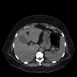
Ring Artefacts
Insufficient exposure results in concentric circles in the center of the field of view.
The ring artifact is seen here in the left lobe of the liver.