Abdominal Aortic Aneurysm
Jump to navigation
Jump to search
Introduction
Case 1
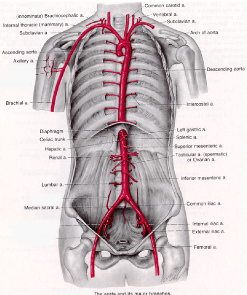
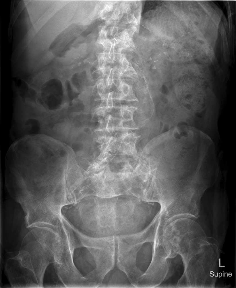
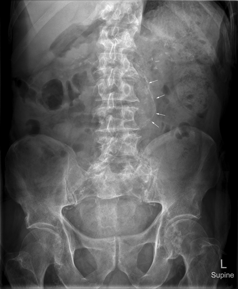
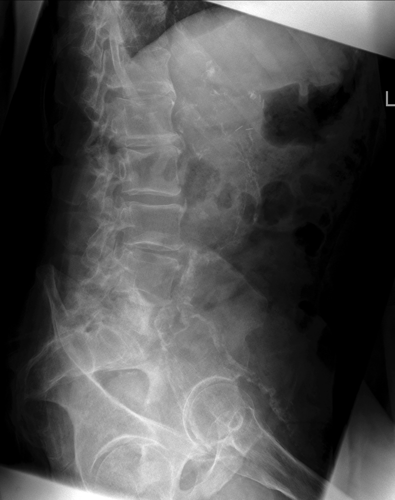
AnatomyAn abdominal aortic aneurysm is commonly referred to as a triple A (AAA). The plain film demonstration of abdominal aortic aneurysm can range in importance from an incidental known finding to a surgical emergency- the distinction is clearly an important one and should be considered by radiographers.
<a class="external" href="http://text.emcrit.org/images/part1/aorta.gif" rel="nofollow" target="_blank">
http://text.emcrit.org/images/part1/aorta.gif (28/2/2011)</a>It begins at the level of the diaphragm, crossing it via the aortic hiatus, technically behind the diaphragm, at the vertebral level of T12. It travels down the posterior wall of the abdomen, anterior to the vertebral column. It thus follows the curvature of the lumbar vertebrae, that is, convex anteriorly. The peak of this convexity is at the level of the third lumbar vertebra (L3). It runs parallel to the inferior vena cava, which is located just to the right of the abdominal aorta, and becomes smaller in diameter as it gives off branches. This is thought to be due to the large size of its principal branches. At the 11th rib, the diameter is about 25 mm; above the origin of the renal arteries, 22 mm; below the renals, 20 mm; and at the bifurcation, 19 mm. (quoted from <a class="external" href="http://en.wikipedia.org/wiki/Abdominal_aorta" rel="nofollow" target="_blank">wikipedia</a>) <a class="external" href="http://www.sutree.com/howto.aspx?from=rss&s=15691" rel="nofollow" target="_blank">Abdominal Aortic Aneurysm
<a class="external" href="http://www.sutree.com/howto.aspx?from=rss&s=15691" rel="nofollow" target="_blank"></a></a><a class="external" href="http://www.sutree.com/howto.aspx?from=rss&s=15691" rel="nofollow" target="_blank">
http://www.sutree.com/howto.aspx?from=rss&s=15691</a> (28/2/2011)
[at Surgery][(28/2/2011)]
Abdominal aortic aneurysm (also known as AAA, pronounced "triple-a") is a localized dilatation (ballooning) of the abdominal aorta exceeding the normal diameter by more than 50 percent, and is the most common form of aortic aneurysm. Approximately 90 percent of abdominal aortic aneurysms occur infrarenally (below the kidneys), but they can also occur pararenally (at the level of the kidneys) or suprarenally (above the kidneys). Such aneurysms can extend to include one or both of the iliac arteries in the pelvis.
Abdominal aortic aneurysms occur most commonly in individuals between 65 and 75 years old and are more common among men and smokers. They tend to cause no symptoms, although occasionally they cause pain in the abdomen and back (due to pressure on surrounding tissues) or in the legs (due to disturbed blood flow). The major complication of abdominal aortic aneurysms is rupture, which can be life-threatening as large amounts of blood spill into the abdominal cavity, and can lead to death within minutes. <a class="external" href="http://en.wikipedia.org/wiki/Abdominal_aortic_aneurysm" rel="nofollow" target="_blank">(quoted from wikipedia)</a>
Relevant Videos
<embed allowfullscreen="true" height="350" src="http://widget.wetpaintserv.us/wiki/wikiradiography/widget/youtubevideo/1e30622af96e464f4d9074984fc84a81101bd59c" type="application/x-shockwave-flash" width="425" wmode="transparent"/> <embed allowfullscreen="true" height="350" src="http://widget.wetpaintserv.us/wiki/wikiradiography/widget/youtubevideo/681ee1f06ab2f75a597846ef8e4ca626a4c3972c" type="application/x-shockwave-flash" width="425" wmode="transparent"/>
Case 1


